
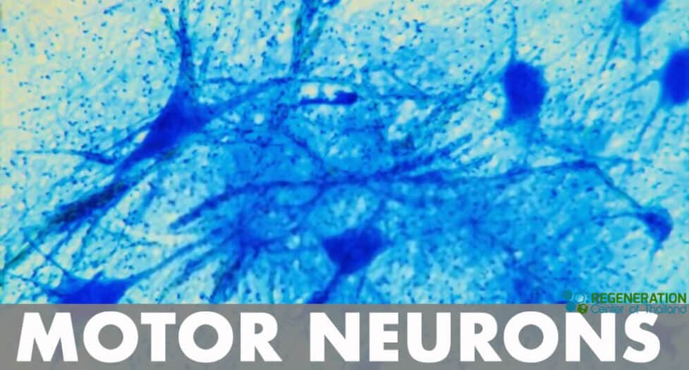
Motoneuron, also known as motor neurons, are a type of cell body located in the brainstem, motor cortex, or spinal cord. Fibers called axons project outwards to the spinal cord can indirectly or directly control muscles, organs, and glands in the body.
The Somatic Motor System
The somatic motor system, also known as the voluntary motor system, is responsible for the movement of skeletal muscles and is under conscious control. It allows for a diverse range of activities, from simple actions like lifting a finger to complex sequences such as playing a musical instrument or participating in athletic events.
- Upper Motor Neurons (UMNs): These are located in the brain’s motor cortex (specifically, the precentral gyrus of the frontal lobe). UMNs send their axons down the spinal cord and synapse with the lower motor neurons. They play a crucial role in initiating voluntary movements and inhibiting involuntary ones.
- Lower Motor Neurons (LMNs): These are located in the spinal cord (anterior horn cells) and the cranial nerve nuclei in the brainstem. LMNs directly innervate skeletal muscles and are responsible for muscle contraction. The axons of LMNs travel out of the central nervous system through spinal nerves (or cranial nerves) and synapse with muscle fibers at neuromuscular junctions.
- Neuromuscular Junction (NMJ): This is the synapse or junction between the axon terminals of LMNs and muscle fibers. At the NMJ, the neurotransmitter acetylcholine is released, leading to muscle contraction.
- Basal Ganglia and Cerebellum: These brain regions are not part of the direct pathway from the motor cortex to the muscles but play vital roles in modulating and refining motor movements. The basal ganglia are involved in the planning, initiation, and regulation of movement, while the cerebellum is essential for coordination, precision, and accurate timing.
- Sensory Feedback: Proprioceptive information, which relates to the body’s position in space and the degree of muscle stretch, provides critical feedback to the central nervous system. This feedback helps in refining motor outputs to achieve the desired movement and posture.
- Motor Planning and Execution: Areas of the brain like the premotor cortex and supplementary motor area contribute to the planning of complex movement sequences. These areas work in conjunction with the primary motor cortex to execute movements.
- Pyramidal and Extrapyramidal Systems: The pyramidal system (consisting of the corticospinal and corticobulbar tracts) is the primary pathway for voluntary movement. The extrapyramidal system, including the basal ganglia and associated pathways, modulates and refines these movements.
Overall, the somatic motor system is a complex and integrated system that allows for the precise control and execution of voluntary movements. Dysfunctions or lesions within this system can lead to a range of motor disorders, such as Parkinson’s disease, Huntington’s disease, spinal cord injuries, Ankylosing Spondylitis or various types of paralysis.
Types of Motor Neurons
- Upper Motor Neurons – Axon fibers from the upper motor neurons help communicate with the spinal cord and other lower motor neuron cells. Another type of
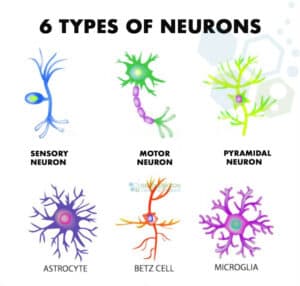 upper motor neurons are pyramidal cells of Betz or Betz cells. These neurotransmitters are known as giant pyramidal cells (neurons) and can be found in the fifth layer of grey matter in the primary motor cortex. These upper motor neurons are the largest cells in the central nervous system and send axon fibers down to the spinal cord along the corticospinal tract. This area is where they connect (synapse) directly with the anterior horn cells and astrocytes.
 - Lower Motor Neurons – Axons in the lower motor neurons help carry signals from our spinal cord motor neurons to muscles or glands (Effectors) and respond to received signals from other motor neurons. There are three types of lower motor neurons, including alpha motor neurons, gamma motor neurons, and beta motor neurons.
- Somatic Motor Neurons (SMN)– SMN starts in the central nervous system and project axons to the skeletal muscles used in locomotion. There are three types of somatic motor neurons including alpha efferent neurons, beta efferent neurons, gamma efferent neurons
- General visceral motor neurons (GVMN) – GVMN help innervate cardiac muscles and the muscles of our arteries). GVMN also connects with ganglia neurons, which are part of the sympathetic and parasympathetic functions in the autonomic nervous system.
- Special visceral motor neurons (SVMN)– SVMN is also known as branchial motor neurons, and help control things such as facial expressions, and swallowing. Learn more about foods that help arthritis.
Why Are Motor Neurons Important?
As seen in the previous example of motor neurons in our brain, the motor nerves primary requirement is the ability to instantly control requires sensory input needed to move accurately. A single motor neuron cell can control thousands of muscle fibers strands, creating innervation to move single muscles (twitch). Motor cell structures in the spinal cord region are considered part of the central nervous system (CNS) and connect to many functions.[1] These motoneurons transmit signals from our spinal cord to the musculoskeletal system for movements. Understanding how to use human neurons in regenerative medicine helps the Regeneration center develop more effective stem cell therapies for patients dealing with spinal cord injuries,Spinal compression fractures, strokes and brain injuries from traumatic events.
What is Innervation?
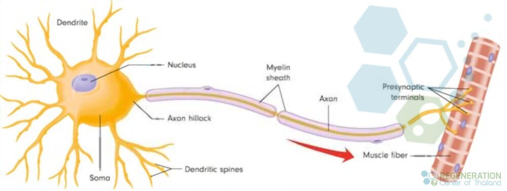
Innervation means “to supply nerves” or “to supply with energy” or “to stimulate.” When nerves embed themselves into muscle fiber, they “innervate” the muscle fibers.[2] This event usually occurs in the neuromuscular junctions and can be identified as simple contractions. They are controlling thousands of individual twitches also us to function and execute movement smoothly and efficiently.
What are Nerve tracts?
Nerve tracts are thick bundles of axons (white matter), these nerve help carry action signals to their effectors. The spinal cord motor nerves carry impulses from many different regions and act as a point of origin for the lower motor cells.[3]
The seven major motor tracts in the spinal cord include: 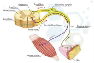
- Anterior corticospinal tract
- Rubrospinal tract
- Lateral corticospinal tract
- Vestibulospinal tract
- Lateral reticulospinal tract
- Tectospinal tract
- Medial reticulospinal tract
What are Dendrites?
Dendrites cells are segments of the neuron that receives stimulation needed neurogenesis and for cells to become activated. Dendrites conduct rapid electrical signals to the neuron cell body asking the cell to function in a specific way. Our nervous system is in charge of the body and controls all other vital system’s functions. Dendrites and neural stem cells help coordinate performance with other systems in the body and are quick to meet our body’s needs in less than one second. The central nervous system uses neuron cells to create movement, and neurons in the brain are necessary for their structure and function. One of the most critical structures in a neuron cell is the dendrite.
Where are cell bodies of somatic motor neurons located?
Neuron cells require stimuli to become active, and Dendrites are the bridge on the neuron that receives the electrical messages. The electric messages come in two primary forms, including excitatory and inhibitory. Excitatory actions and paracrine cell signalling increase the stimulation of a neuron cell, and inhibitory actions help decrease a neuron’s activity. These signals are accumulated in the neuron cell body (soma) after being collected by dendrites. Once dendrites receive these action signals, they will get sent to an area in the soma called the axon hillock. This area would be in the neck part of a cell body. Once neurons receive enough excitatory signals, they become activated and proceed to create more action potential for other cells.[4]
Damaged Neurons & Dendrites in Neurodegenerative disease
Neurodegenerative disorders are generally characterized as progressive loss of the function and structure neuron cells associated with neuronal cell death (apoptosis). The most common neurodegenerative disorders include: Motor Neuron Disease (MND), Ataxia, Multiple sclerosis , Parkinson’s disease (PD), Alzheimer’s disease (AD), and amyotrophic lateral sclerosis (ALS).
Published Clinical Citations
[1] ^ Benkler, Chen, Alison L O’Neil, Susannah Slepian, Fang Qian, Paul H Weinreb, and Lee L Rubin. 2018. Aggregated SOD1 causes selective death of cultured human motor neurons. Scientific reports, no. 1 (November 6). doi:10.1038/s41598-018-34759-z. https://www.ncbi.nlm.nih.gov/pubmed/30401824
[2] ^ Holzbaur, Erika L F. 2004. Motor neurons rely on motor proteins. Trends in cell biology, no. 5. https://www.ncbi.nlm.nih.gov/pubmed/15130579
[3] ^ Sellers, Drew L, Jamie M Bergen, Russell N Johnson, Heidi Back, John M Ravits, Philip J Horner, and Suzie H Pun. 2016. Targeted axonal import (TAxI) peptide delivers functional proteins into spinal cord motor neurons after peripheral administration. Proceedings of the National Academy of Sciences of the United States of America, no. 9 (February 17). doi:10.1073/pnas.1515526113. https://www.ncbi.nlm.nih.gov/pubmed/26888285
[4] ^ Kim, Minkyung, Brielle Bjorke, and Grant S Mastick. 2017. Motor neuron migration and positioning mechanisms: New roles for guidance cues. Seminars in cell & developmental biology (November 14). doi:S1084-9521(17)30465-2. https://www.ncbi.nlm.nih.gov/pubmed/29141180