Neurons, or nerve cells, are the signalling cells of the nervous system. Each one receives signals on its dendrites and cell body, decides whether to fire, and sends an electrical impulse, the action potential, along its axon. At the far end, at a junction called a synapse, the electrical signal is turned into a chemical one: the neuron releases a neurotransmitter that acts on the next cell. The adult human brain contains about 86 billion neurons.[1] This page covers how a neuron is built, how fast it signals, and what the main neurotransmitters do.
Overview of neurons:
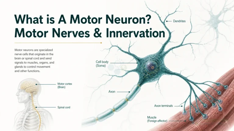
- Cell body (soma). Contains the nucleus and most of the cell’s protein-making machinery.
- Dendrites. Usually highly branched processes that receive information from other neurons at synapses. Information flows from the dendrites, across the cell body and down the axon.[2]
- Axon hillock and initial segment. Where the axon leaves the cell body. Together they form the trigger zone, where action potentials begin.[2]
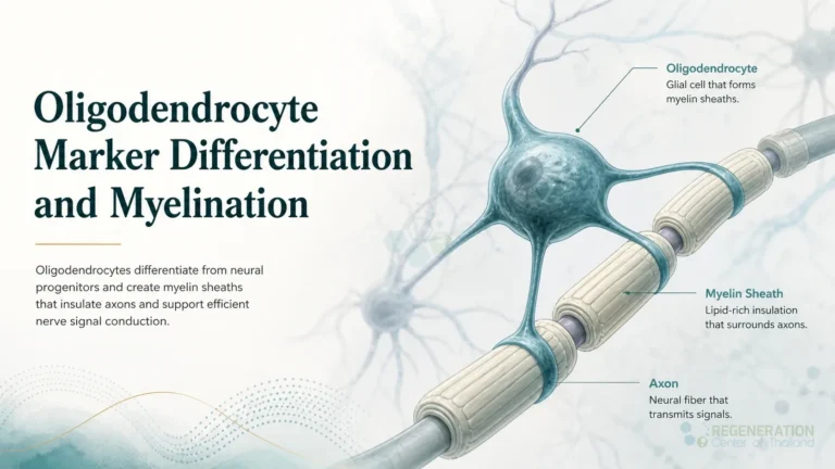
- Axon. The single long fibre that carries the impulse away from the cell body. Many axons are wrapped in myelin, an insulating sheath made by glial cells: oligodendrocytes in the brain and spinal cord, Schwann cells in peripheral nerves. The short unmyelinated gaps between myelin segments are the nodes of Ranvier.[2]
- Axon terminals. Branches at the end of the axon, each ending in a synaptic end bulb that makes the connection with the target cell at the synapse.[2]
Neurons are classified by shape and by job. By shape, most are multipolar, with one axon and two or more dendrites; bipolar neurons, with one axon and one dendrite, are found mainly in the retina and the olfactory epithelium; and the sensory neurons whose cell bodies sit in ganglia outside the brain and spinal cord are pseudo-unipolar, with a single process that splits into two branches.[2] By job, sensory (afferent) neurons carry information into the central nervous system, motor (efferent) neurons carry commands out to muscles and glands, and interneurons connect neurons within the brain and spinal cord. Motor neurons have their own page on somatic motor neurons and innervation.
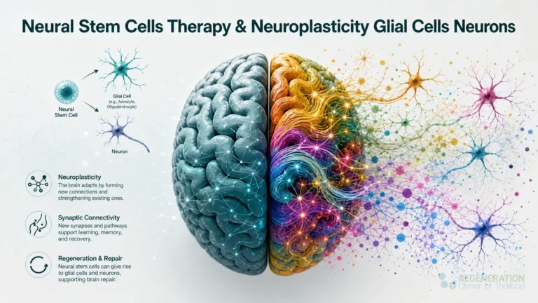
Neurons depend on glial cells, which insulate axons, keep the fluid around synapses stable and clear away debris. Glial cells, and the neural stem cells that produce both neurons and glia, are described on our page on neural stem cells and glial cells.
Anatomy of a Neuron – VIDEO
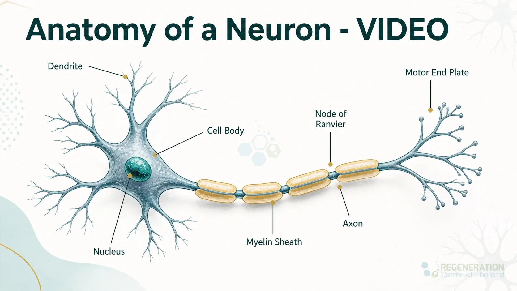
The diagram shows a myelinated neuron. In a motor neuron, the axon terminals end on muscle fibres, and the muscle side of that junction is called the motor end plate. Neurons in the adult brain are long-lived cells; most are never replaced during life or after injury.[3] Whether the adult human hippocampus keeps adding a small number of new neurons is still debated, and the evidence on both sides is set out on our page on neurogenesis. Injury, stroke and diseases such as Alzheimer’s disease and ALS have different causes, but each damages neurons or the connections between them.
What is the speed of neural signaling?
At rest, the inside of a neuron is about 70 millivolts negative compared with the outside. When incoming signals push the trigger zone to about -55 mV, the threshold, voltage-gated sodium channels open, sodium rushes in and the membrane swings to about +30 mV; potassium channels then open and return it to rest. This brief reversal, the action potential, is regenerated patch by patch along the axon.[4]
How fast it travels depends on two things: the diameter of the axon, because current spreads faster down a wide axon than a narrow one, and myelin.[4] In an unmyelinated axon the impulse moves by continuous conduction, opening channels all along its length, which is slow. In a myelinated axon the voltage-gated channels are concentrated at the nodes of Ranvier, and the impulse effectively jumps from node to node. This is called saltatory conduction, from the Latin saltare, to leap, and it is both faster and more energy-efficient.[4][5]
The result is a range of speeds of more than a hundredfold. A standard classification of human peripheral nerve fibres gives these values:[6]
| Fibre type | Diameter | Conduction velocity | Notes |
|---|---|---|---|
| A-alpha (myelinated) | 12 to 22 µm | 70 to 120 m/s | Thick myelinated fibres; in motor nerves, the alpha fibres supply ordinary (extrafusal) muscle fibres |
| A-beta (myelinated) | 5 to 12 µm | 30 to 70 m/s | Thick myelinated fibres |
| A-gamma (myelinated) | 2 to 8 µm | 15 to 30 m/s | In motor nerves, the gamma fibres supply the muscle fibres inside muscle spindles |
| A-delta (thinly myelinated) | 1 to 5 µm | 5 to 30 m/s | Thin myelinated fibres, intermediate speed |
| B (myelinated) | Under 3 µm | 3 to 15 m/s | Sympathetic preganglionic fibres |
| C (unmyelinated) | 0.1 to 1.3 µm | 0.6 to 2.0 m/s | Unmyelinated fibres, the slowest |
At 100 m/s, a signal crosses a metre of nerve in 10 milliseconds; at 1 m/s, the same distance takes a full second. When myelin is damaged, as in multiple sclerosis, current leaks from the previously insulated stretches of axon and conduction slows or fails.[5] How the oligodendrocytes that make central myelin develop is covered on our page on oligodendrocyte markers and myelination.
What are Neurotransmitters?
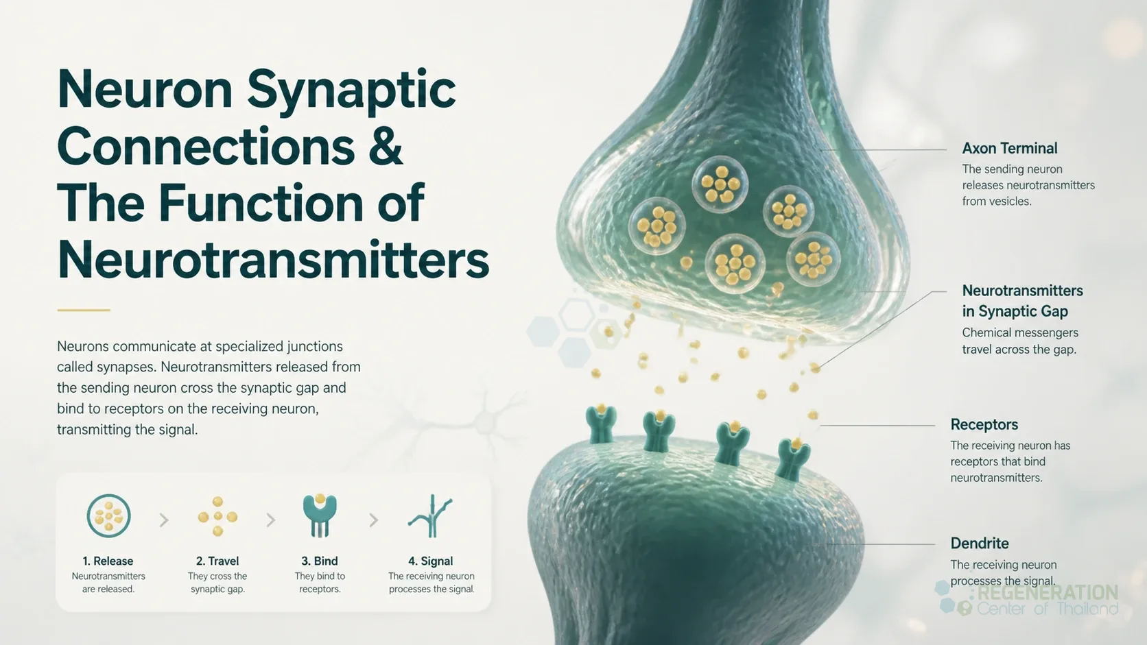
Neurotransmitters are the chemical messengers that carry a signal across the synapse, from a neuron to another neuron, a muscle cell or a gland cell. Every chemical synapse has the same parts: a presynaptic terminal holding the neurotransmitter packed in vesicles, a narrow gap called the synaptic cleft, receptor proteins on the postsynaptic cell, and a way of removing the transmitter afterwards.[7]
Transmission runs in a fixed order. When an action potential reaches the axon terminal, voltage-gated calcium channels open and calcium flows in. Calcium makes the vesicles fuse with the membrane and release their transmitter into the cleft. The transmitter binds its receptors on the other side and is then cleared, either broken down by an enzyme, as acetylcholinesterase breaks down acetylcholine, or taken back up by the neuron or a neighbouring glial cell to be recycled.[7] Neurotransmission is one of several ways cells signal to each other; others are covered on our page on paracrine signaling.
What is the function of neurotransmitters and how do they affect neurons?
Whether a neurotransmitter excites or inhibits depends mainly on the receptor it binds, not on the molecule alone.[7] An excitatory effect depolarizes the receiving neuron, moving it closer to firing; an inhibitory effect hyperpolarizes it, moving it further away. Each neuron adds up many such inputs, and fires only if the sum reaches threshold at the trigger zone.
- Ionotropic receptors are ion channels that open when the transmitter binds, so they act within milliseconds. The nicotinic acetylcholine receptor at the muscle junction is an example.[7]
- Metabotropic receptors work through a G protein and an effector protein inside the cell. They act more slowly and can change the cell’s metabolism or gene activity rather than its voltage alone.[7]
The same transmitter can do opposite things at different receptors. Acetylcholine excites skeletal muscle through nicotinic receptors but can excite or inhibit through its muscarinic receptors, and dopamine’s D1 receptors are excitatory while its D2 receptors are inhibitory.[7] Transmitters that act mostly through slower metabotropic receptors and adjust the activity of whole groups of neurons, such as dopamine, serotonin and noradrenaline, are often called neuromodulators.
Types of Neurotransmitters
Neurotransmitters fall into four main chemical groups: acetylcholine, which forms its own group; amino acids (glutamate, GABA and glycine); biogenic amines made from amino acids (serotonin from tryptophan; dopamine, noradrenaline and adrenaline from tyrosine); and neuropeptides, short chains of amino acids such as the five-amino-acid met-enkephalin and the 31-amino-acid beta-endorphin, which are often released together with another transmitter.[7] The main ones:
| Neurotransmitter | Group | Usual effect | Main roles |
|---|---|---|---|
| Glutamate | Amino acid | Excitatory | The main excitatory transmitter of the adult brain; learning and memory[8][9] |
| GABA (gamma-aminobutyric acid) | Amino acid | Inhibitory | Inhibitory signalling in the brain; involved in brain function and sleep, with effects that include reduced anxiety and tension[7][9] |
| Glycine | Amino acid | Inhibitory | Inhibitory signalling in the central nervous system[7] |
| Acetylcholine | Acetylcholine | Excitatory at muscle; mixed elsewhere | Triggers contraction at the neuromuscular junction; also used in the autonomic nervous system and throughout the brain, where it is involved in memory[7][9] |
| Dopamine | Biogenic amine | Depends on receptor (D1 or D2) | Mood, sleep and learning, with effects that include pleasure; low dopamine in Parkinson’s disease, overactive dopamine signalling linked to some symptoms of schizophrenia[9] |
| Serotonin (5-HT) | Biogenic amine | Depends on receptor | Mood, sleep and appetite in the brain; most of the body’s serotonin is made in the gut[9][10] |
| Noradrenaline (norepinephrine) | Biogenic amine | Depends on receptor (alpha or beta) | Alertness and arousal; heart and gut activity; also released into the blood as a hormone by the adrenal gland[7][9] |
Labels such as “the pleasure chemical” or “the mood chemical” are simplifications. Each transmitter acts through several receptor types in many brain circuits, and most behaviours involve several transmitters at once.
Can excitatory neurochemicals affect mood & motivation?
Yes: both too little and too much signalling can cause problems. Dopamine is involved in mood and learning, and its effects include pleasure; drugs that block or mimic transmitters change behaviour: dopamine agonists, which mimic dopamine at its receptors, are a common treatment strategy for Parkinson’s disease, and the antipsychotic drugs used for some symptoms of schizophrenia block dopamine receptors.[9]
Too much excitation is harmful. During a stroke, the concentration of glutamate outside neurons rises abruptly, and overstimulation of NMDA-type glutamate receptors triggers a chain of events that kills neurons, a process called excitotoxicity. This finding led to clinical trials of drugs that block excitotoxicity in stroke patients, but the first generation of these drugs failed.[8]
The link between neurotransmitters and mood is less direct than the popular “chemical imbalance” picture suggests. A 2023 umbrella review of the main lines of research, including serotonin levels, receptor and transporter imaging, tryptophan depletion and genetics, found no consistent evidence that depression is associated with lower serotonin concentration or activity.[11] Other researchers have disputed that conclusion, pointing to methodological weaknesses and to evidence from tryptophan depletion and serotonin imaging studies.[12] The debate is about the cause of depression, not about treatment: in a network meta-analysis of 522 double-blind trials with 116,477 adults with major depressive disorder, all 21 antidepressants studied were more effective than placebo in acute treatment.[13] Questions about any medicine are best discussed with the prescribing doctor.
Are There Neurotransmitters in the Gut?
Yes. The gut has its own nervous system, the enteric nervous system, which controls the muscles of the digestive tract and the movement of fluid across its lining, works together with signals from the brain and spinal cord, and uses a wide range of chemical messengers.[14]
Serotonin is the clearest example. More than 90% of the body’s serotonin is made in the gut, mainly by enterochromaffin cells in its lining.[10] These cells act as sensors: their serotonin activates nerve fibres in the gut wall that start the reflexes of peristalsis and secretion, and others that carry information to the central nervous system. Enterochromaffin cells make serotonin with the enzyme tryptophan hydroxylase 1, while neurons, including those in the gut wall, use tryptophan hydroxylase 2.[15] Gut serotonin is taken up by blood platelets and helps regulate gut movement, blood clotting, immune responses and, in animal studies, bone development; in mice, spore-forming bacteria of the normal gut flora raise its production.[10] Serotonin in the brain is made by the brain’s own neurons,[15] and the brain sits behind the blood-brain barrier, through which very little passes by simple diffusion.[2]
Several common medicines work on gut serotonin receptors. Ondansetron and granisetron, which block 5-HT3 receptors, are used against the nausea of cancer chemotherapy, and alosetron, another 5-HT3 blocker, is used for irritable bowel syndrome with diarrhoea.[15]
Making Neurons in the Laboratory
Neurons can now be grown from stem cells. In one method, human embryonic or induced pluripotent stem cells were turned into neural stem cells in seven days; these could be expanded and then made into neurons, astrocytes and oligodendrocytes, including GABA-releasing, dopamine-releasing and motor neurons.[16] When cortical neurons made from human induced pluripotent stem cells were transplanted into the stroke-damaged cortex of rats, they matured into functional neurons, and the treated rats recovered better on a movement test, although at that early point the improvement was most likely not due to the new neurons rewiring the brain.[17] What transplanted neural cells have and have not achieved in human trials is summarized on our page on neural stem cells.
Published Clinical Citations
[1] ^ Azevedo FA, Carvalho LR, Grinberg LT, et al. Equal numbers of neuronal and nonneuronal cells make the human brain an isometrically scaled-up primate brain. Journal of Comparative Neurology. 2009;513(5):532-541. https://doi.org/10.1002/cne.21974
[2] ^ Betts JG, Young KA, Wise JA, et al. Anatomy and Physiology 2e. Section 12.2, Nervous Tissue. OpenStax, Rice University; 2022. https://openstax.org/books/anatomy-and-physiology-2e/pages/12-2-nervous-tissue
[3] ^ Magnusson JP, Göritz C, Tatarishvili J, et al. A latent neurogenic program in astrocytes regulated by Notch signaling in the mouse. Science. 2014;346(6206):237-241. https://doi.org/10.1126/science.346.6206.237
[4] ^ Betts JG, Young KA, Wise JA, et al. Anatomy and Physiology 2e. Section 12.4, The Action Potential. OpenStax, Rice University; 2022. https://openstax.org/books/anatomy-and-physiology-2e/pages/12-4-the-action-potential
[5] ^ Clark MA, Douglas M, Choi J. Biology 2e. Section 35.2, How Neurons Communicate. OpenStax, Rice University; 2018. https://openstax.org/books/biology-2e/pages/35-2-how-neurons-communicate
[6] ^ Manzano GM, Giuliano LM, Nóbrega JA. A brief historical note on the classification of nerve fibers. Arquivos de Neuro-Psiquiatria. 2008;66(1):117-119. https://doi.org/10.1590/s0004-282×2008000100033
[7] ^ Betts JG, Young KA, Wise JA, et al. Anatomy and Physiology 2e. Section 12.5, Communication Between Neurons. OpenStax, Rice University; 2022. https://openstax.org/books/anatomy-and-physiology-2e/pages/12-5-communication-between-neurons
[8] ^ Lai TW, Zhang S, Wang YT. Excitotoxicity and stroke: identifying novel targets for neuroprotection. Progress in Neurobiology. 2014;115:157-188. https://doi.org/10.1016/j.pneurobio.2013.11.006
[9] ^ Spielman RM, Jenkins WJ, Lovett MD. Psychology 2e. Section 3.2, Cells of the Nervous System. OpenStax, Rice University; 2020. https://openstax.org/books/psychology-2e/pages/3-2-cells-of-the-nervous-system
[10] ^ Yano JM, Yu K, Donaldson GP, et al. Indigenous bacteria from the gut microbiota regulate host serotonin biosynthesis. Cell. 2015;161(2):264-276. https://doi.org/10.1016/j.cell.2015.02.047
[11] ^ Moncrieff J, Cooper RE, Stockmann T, et al. The serotonin theory of depression: a systematic umbrella review of the evidence. Molecular Psychiatry. 2023;28(8):3243-3256. https://doi.org/10.1038/s41380-022-01661-0
[12] ^ Jauhar S, Arnone D, Baldwin DS, et al. A leaky umbrella has little value: evidence clearly indicates the serotonin system is implicated in depression. Molecular Psychiatry. 2023;28(8):3149-3152. https://doi.org/10.1038/s41380-023-02095-y
[13] ^ Cipriani A, Furukawa TA, Salanti G, et al. Comparative efficacy and acceptability of 21 antidepressant drugs for the acute treatment of adults with major depressive disorder: a systematic review and network meta-analysis. Lancet. 2018;391(10128):1357-1366. https://doi.org/10.1016/S0140-6736(17)32802-7
[14] ^ Furness JB. The enteric nervous system and neurogastroenterology. Nature Reviews Gastroenterology & Hepatology. 2012;9(5):286-294. https://doi.org/10.1038/nrgastro.2012.32
[15] ^ Gershon MD, Tack J. The serotonin signaling system: from basic understanding to drug development for functional GI disorders. Gastroenterology. 2007;132(1):397-414. https://doi.org/10.1053/j.gastro.2006.11.002
[16] ^ Yan Y, Shin S, Jha BS, et al. Efficient and rapid derivation of primitive neural stem cells and generation of brain subtype neurons from human pluripotent stem cells. Stem Cells Translational Medicine. 2013;2(11):862-870. https://doi.org/10.5966/sctm.2013-0080
[17] ^ Tornero D, Wattananit S, Grønning Madsen M, et al. Human induced pluripotent stem cell-derived cortical neurons integrate in stroke-injured cortex and improve functional recovery. Brain. 2013;136(Pt 12):3561-3577. https://doi.org/10.1093/brain/awt278