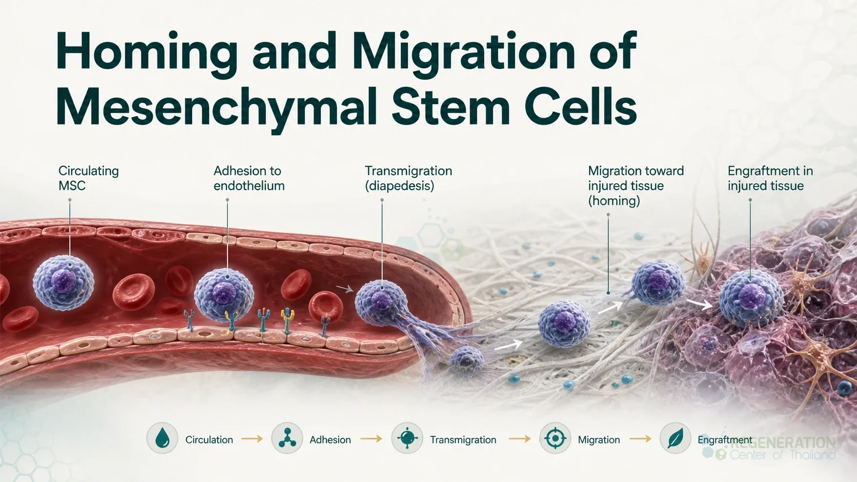
Homing is the journey a transplanted or infused stem cell makes from the bloodstream into a tissue, ending with the cell settling in a particular place. It is best understood for blood-forming (hematopoietic) stem cells reaching the bone marrow after a transplant, where it is the first and essential step.[1] For the mesenchymal stromal cells (MSCs) used in many experimental cell treatments, most infused cells never reach the target tissue: after injection into a vein they are caught in the lungs first, and few survive long. What is known about where they go, and the signalling and adhesion steps involved, is set out below. The molecules that direct the movement have their own page on chemokines.
The Multistep Homing Model
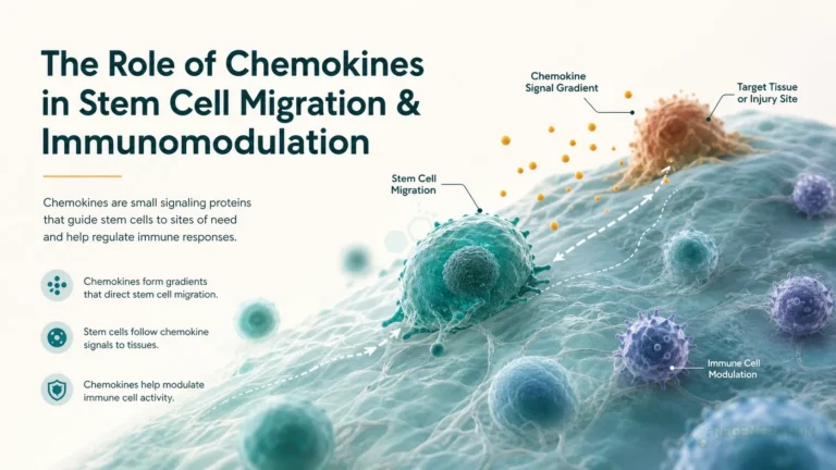
Homing follows the same sequence of steps that white blood cells use to leave the circulation. For blood-forming stem cells reaching the marrow it involves signalling through stromal cell-derived factor 1 (SDF-1, also called CXCL12) and stem cell factor, activation of the adhesion molecules LFA-1, VLA-4, VLA-5 and CD44, rearrangement of the cell’s internal skeleton, and the enzymes MT1-MMP and MMP-2 and MMP-9. Rolling and firm adhesion to the lining cells of the small marrow blood vessels under flowing blood is followed by migration across that lining and the matrix beneath it, and finally by anchoring in a niche next to the bone surface or around blood vessels.[1]
| Step | What happens | Molecules involved |
|---|---|---|
| 1. Tethering and rolling | The flowing cell makes brief contacts with the vessel lining and slows down | Selectins on the lining cells. MSCs rolled on human endothelial cells in a flow chamber, and the rolling was blocked by an antibody against P-selectin; MSCs also rolled on immobilised P-selectin.[2] MSCs lack PSGL-1, the main white blood cell selectin ligand, so how they bind P-selectin is not settled[3] |
| 2. Activation by chemokines | Chemokines on the vessel surface switch the cell’s adhesion molecules into a sticky state | SDF-1 and its receptor CXCR4.[1] Only a small subset of cultured human MSCs carries working CXCR4; in laboratory migration tests the receptor was important for their movement toward bone marrow[4] |
| 3. Firm adhesion | The cell stops and holds on to the vessel wall | Integrin VLA-4 binding VCAM-1; blocking either stopped MSCs binding to endothelial cells[2] |
| 4. Transmigration | The cell squeezes between or through the lining cells into the tissue | MSCs cross mainly between lining cells, more slowly than white blood cells[3] |
| 5. Invasion of the matrix | The cell digests a path through the basement membrane and matrix | MMP-2, MT1-MMP and TIMP-2; silencing any of them cut the ability of human MSCs to cross a reconstituted basement membrane, and inflammatory signals increased it[5] |
| 6. Lodging in a niche | The cell anchors in a particular microenvironment | For blood-forming stem cells, niches at the bone surface and around vessels[1] |
The model comes mainly from blood-forming cells and white blood cells, and MSCs do not follow it exactly: they are considerably larger than white blood cells and some of their arrest in small vessels is passive rather than driven by adhesion molecules.[3] Cells can be engineered to home better. Adding a sugar group to the surface protein CD44 of human MSCs in the laboratory created a binding site for E-selectin, and the treated cells entered the marrow of mice within hours of infusion.[6] Growing mouse bone marrow cells in culture raised their CXCR4 and their migration toward SDF-1; when injected into the leg muscle of mice with a blocked leg artery, the cultured cells stayed in the tissue and restored more blood flow than fresh cells.[7]
Matrix metalloproteinases help migrating cells cut a path through the basement membrane and matrix,[5] and they also trim signalling proteins, making some chemokines more active and inactivating others, such as SDF-1 (CXCL12).[8]
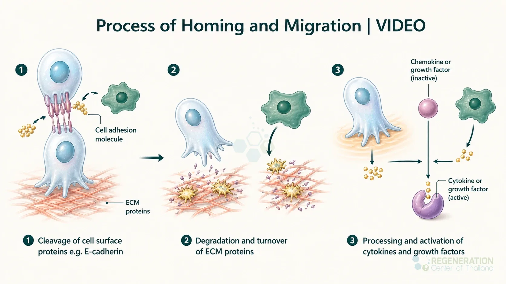
Process of Homing and Migration | VIDEO
The animation shows the same cascade for a neutrophil, a white blood cell, following a chemotactic gradient from a site of injury.
Where Infused Cells Actually Go
This is the practical point for anyone considering a cell treatment given into a vein.
- First-pass lung trapping. In rats, most MSCs infused into a vein were trapped in the lungs; fewer reached the arterial circulation than with smaller cell types, and blocking the adhesion molecule CD49d increased the number that passed through.[9] Mouse MSCs measured 15 to 19 micrometres across in suspension, and within seconds of injection the great majority were in the lungs.[10]
- Short survival. In mice, two million human MSCs given into a vein were trapped as clumps in the lungs and disappeared with a half-life of about 24 hours; fewer than 1,000 cells were found in six other tissues. The cells still reduced inflammation and heart attack size, through the anti-inflammatory protein TSG-6 they released in the lungs.[11]
- Human imaging. In four patients with liver cirrhosis, MSCs labelled with indium-111 and infused into a vein were seen first in the lungs; over 10 days the signal in the lungs fell from 27 to 33 percent of the injected activity down to 2 to 5 percent, while the liver rose to 13 to 17 percent and the spleen to 30 to 42 percent.[12]
- Delivery route matters. In nine patients after a heart attack, three in each group, bone marrow cells labelled with a radioactive sugar and given into the coronary artery left 1.3 to 2.6 percent of the activity in the damaged heart muscle 50 to 75 minutes later, with the rest mainly in liver and spleen; after infusion into an arm vein, only background activity was seen in the heart. Cells enriched for the marker CD34 and given into the artery left 14 to 39 percent.[13]
- Long-term engraftment is limited. In autopsy tissue from 18 patients who had received donor MSCs, no tissue or tumour of donor origin was found, and donor DNA was detectable at low levels (1 in 100 to fewer than 1 in 1,000 cells) mainly when the infusion had been given within the previous 50 days.[14]
Across experimental and clinical studies using imaging, the share of infused cells that reaches the target tissue has been low.[15] This is why the effects of MSC treatments are now attributed mainly to the signals the cells release while they are in the body rather than to their replacing damaged tissue, an argument set out on our pages on paracrine signaling and the secretome. Infused MSCs are also taken up by the recipient’s own immune cells: in mice, dying MSCs were engulfed by monocytes in the lungs and liver, and those monocytes then changed behaviour and travelled elsewhere in the body.[16] More on these effects is on our page on immunomodulation.
What Changes Homing
Reported influences on how many cells arrive include the number of times the cells have been grown and split in culture, the growth medium (see cell culture growth medium), the dose and the delivery route, the recipient’s age, and how much inflammation there is in the target tissue, since inflammatory signals raise the chemokines and adhesion molecules on the vessel lining.[15][5] Cells can be given into a vein, into an artery supplying the organ, directly into the tissue, into a joint, or into the fluid around the spinal cord (see intrathecal administration); the more local routes avoid the lungs but are more invasive.
Mobilization: The Journey in Reverse
Blood-forming stem cells live in the bone marrow but can be released into the bloodstream, which is called mobilization. In practice this is done with medicines such as G-CSF so that large numbers of these somatic stem cells can be collected from the blood by apheresis for a hematopoietic cell transplant.[17] Homing during normal life also has a role in the body’s day-to-day balance, and it increases when stress recruits white blood cells from the marrow reserve.[1] Drugs that block CXCR4, such as plerixafor, are added to G-CSF to release more of these cells; they are described on our page on chemokines.[17]
Published Clinical Citations
[1] ^ Lapidot T, Dar A, Kollet O. How do stem cells find their way home? Blood. 2005;106(6):1901-1910. https://doi.org/10.1182/blood-2005-04-1417
[2] ^ Rüster B, Göttig S, Ludwig RJ, et al. Mesenchymal stem cells display coordinated rolling and adhesion behavior on endothelial cells. Blood. 2006;108(12):3938-3944. https://doi.org/10.1182/blood-2006-05-025098
[3] ^ Nitzsche F, Müller C, Lukomska B, Jolkkonen J, Deten A, Boltze J. Concise review: MSC adhesion cascade, insights into homing and transendothelial migration. Stem Cells. 2017;35(6):1446-1460. https://doi.org/10.1002/stem.2614
[4] ^ Wynn RF, Hart CA, Corradi-Perini C, et al. A small proportion of mesenchymal stem cells strongly expresses functionally active CXCR4 receptor capable of promoting migration to bone marrow. Blood. 2004;104(9):2643-2645. https://doi.org/10.1182/blood-2004-02-0526
[5] ^ Ries C, Egea V, Karow M, Kolb H, Jochum M, Neth P. MMP-2, MT1-MMP, and TIMP-2 are essential for the invasive capacity of human mesenchymal stem cells: differential regulation by inflammatory cytokines. Blood. 2007;109(9):4055-4063. https://doi.org/10.1182/blood-2006-10-051060
[6] ^ Sackstein R, Merzaban JS, Cain DW, et al. Ex vivo glycan engineering of CD44 programs human multipotent mesenchymal stromal cell trafficking to bone. Nature Medicine. 2008;14(2):181-187. https://doi.org/10.1038/nm1703
[7] ^ Shiba Y, Takahashi M, Hata T, et al. Bone marrow CXCR4 induction by cultivation enhances therapeutic angiogenesis. Cardiovascular Research. 2009;81(1):169-177. https://doi.org/10.1093/cvr/cvn247
[8] ^ Hughes CE, Nibbs RJB. A guide to chemokines and their receptors. FEBS Journal. 2018;285(16):2944-2971. https://doi.org/10.1111/febs.14466
[9] ^ Fischer UM, Harting MT, Jimenez F, et al. Pulmonary passage is a major obstacle for intravenous stem cell delivery: the pulmonary first-pass effect. Stem Cells and Development. 2009;18(5):683-692. https://doi.org/10.1089/scd.2008.0253
[10] ^ Schrepfer S, Deuse T, Reichenspurner H, Fischbein MP, Robbins RC, Pelletier MP. Stem cell transplantation: the lung barrier. Transplantation Proceedings. 2007;39(2):573-576. https://doi.org/10.1016/j.transproceed.2006.12.019
[11] ^ Lee RH, Pulin AA, Seo MJ, et al. Intravenous hMSCs improve myocardial infarction in mice because cells embolized in lung are activated to secrete the anti-inflammatory protein TSG-6. Cell Stem Cell. 2009;5(1):54-63. https://doi.org/10.1016/j.stem.2009.05.003
[12] ^ Gholamrezanezhad A, Mirpour S, Bagheri M, et al. In vivo tracking of 111In-oxine labeled mesenchymal stem cells following infusion in patients with advanced cirrhosis. Nuclear Medicine and Biology. 2011;38(7):961-967. https://doi.org/10.1016/j.nucmedbio.2011.03.008
[13] ^ Hofmann M, Wollert KC, Meyer GP, et al. Monitoring of bone marrow cell homing into the infarcted human myocardium. Circulation. 2005;111(17):2198-2202. https://doi.org/10.1161/01.CIR.0000163546.27639.AA
[14] ^ von Bahr L, Batsis I, Moll G, et al. Analysis of tissues following mesenchymal stromal cell therapy in humans indicates limited long-term engraftment and no ectopic tissue formation. Stem Cells. 2012;30(7):1575-1578. https://doi.org/10.1002/stem.1118
[15] ^ Chavakis E, Urbich C, Dimmeler S. Homing and engraftment of progenitor cells: a prerequisite for cell therapy. Journal of Molecular and Cellular Cardiology. 2008;45(4):514-522. https://doi.org/10.1016/j.yjmcc.2008.01.004
[16] ^ de Witte SFH, Luk F, Sierra Parraga JM, et al. Immunomodulation by therapeutic mesenchymal stromal cells (MSC) is triggered through phagocytosis of MSC by monocytic cells. Stem Cells. 2018;36(4):602-615. https://doi.org/10.1002/stem.2779
[17] ^ Sanofi-aventis US LLC. MOZOBIL (plerixafor) injection, for subcutaneous use. US prescribing information. DailyMed set ID 0ed08d2b-5051-46b2-aa37-1d6275bf9003. https://dailymed.nlm.nih.gov/dailymed/drugInfo.cfm?setid=0ed08d2b-5051-46b2-aa37-1d6275bf9003