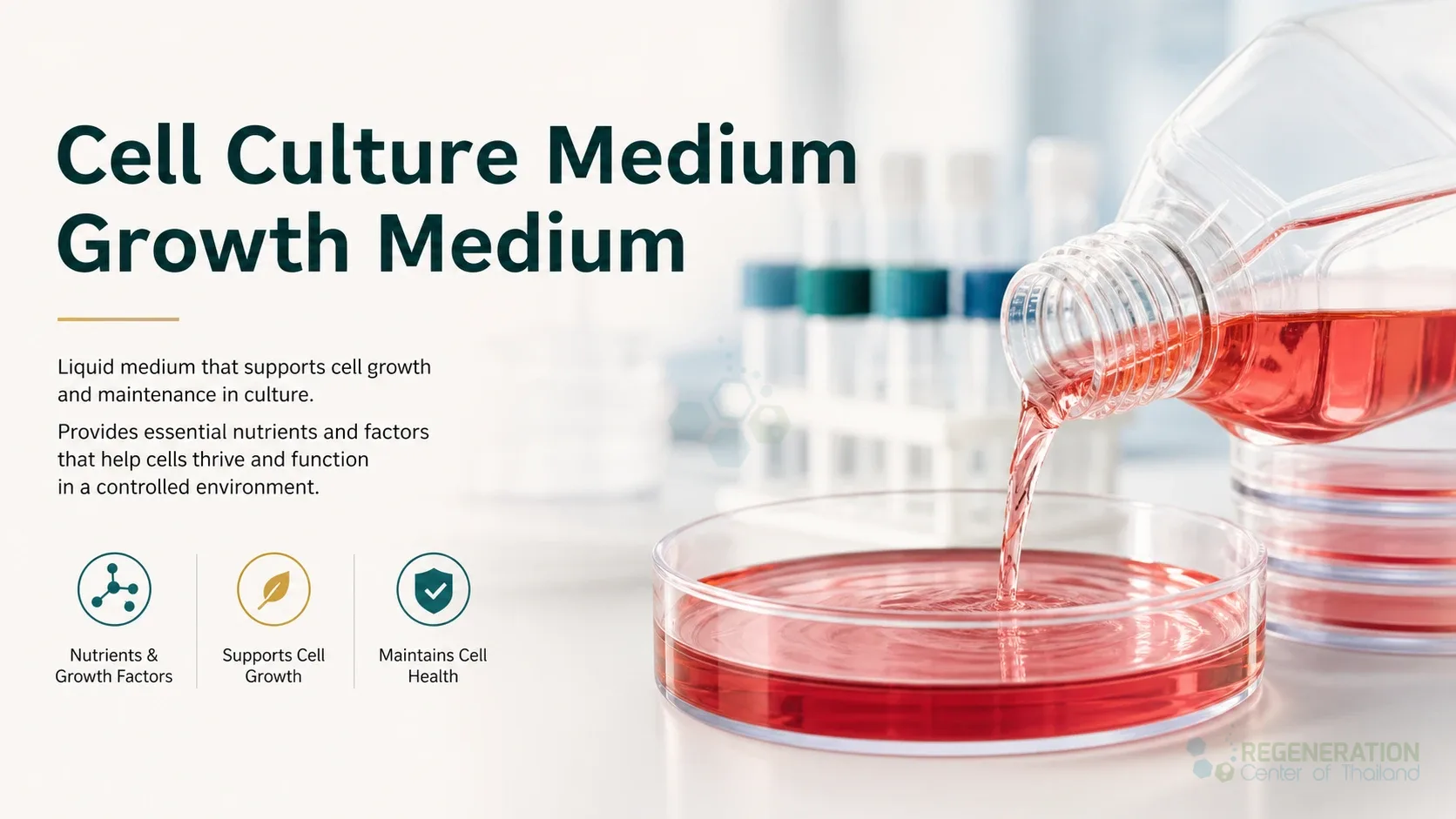
Cell culture medium, also called growth medium, is the liquid in which cells are kept alive and allowed to multiply outside the body. It has to do the work that blood and tissue fluid do inside the body: supply energy and the building blocks for proteins and DNA, keep the salt balance and acidity (pH) within a narrow range, and, when the cells need them, carry the hormones and growth factors that signal them to divide or to specialize. Most media in use today have two parts: a basal medium, which is a fixed mixture of salts, sugar, amino acids and vitamins, and supplements added to it, such as serum or a defined set of proteins.[1]
This page explains what goes into a medium and why each part is there. How cells are taken from tissue, grown, passaged and tested, and how cells meant for patients are made under good manufacturing practice, is covered on our page on human cell culturing.
Components of Cell Culture Medium:
| Component | Why it is there |
|---|---|
| Inorganic salts (sodium, potassium, calcium, magnesium, phosphate) | Set the osmotic pressure and ion levels close to those of body fluids. Early media were simple balanced salt solutions of this kind, which keep tissue alive for only a few days.[1] |
| Glucose | A source of energy. Breaking down glucose produces lactic acid, so fast-growing cells in a high-glucose medium build up lactate and make the medium more acidic.[1] |
| Amino acids | Building blocks for proteins. Essential amino acids must be supplied; non-essential ones can be made by the cell, but adding them lightens its load. Glutamine is also an energy source, but it breaks down in the medium into ammonia, which is toxic to cells, so it is added shortly before use or replaced by a more stable form such as L-alanyl-L-glutamine.[1] |
| Vitamins | Needed in small amounts for cell metabolism. Basal media differ in which vitamins they contain: Eagle’s basal medium of 1955 had eight, while others, such as Medium 199, also include the fat-soluble vitamins.[1] |
| Buffer | Keeps the pH stable. Most media rely on sodium bicarbonate balanced against the carbon dioxide in the incubator; HEPES is sometimes added for extra buffering, but it can generate hydrogen peroxide when the medium is exposed to light.[1] |
| pH indicator | Phenol red changes color as the medium turns acidic. It can affect the growth of some cells and has estrogen-like activity, which matters for hormone-sensitive cells such as breast tissue cells.[1] |
| Serum | A source of hormones, growth factors, carrier proteins and lipids, which is why it supports so many cell types. Its exact composition is unknown and varies from batch to batch.[1] |
| Defined supplements | Purified or recombinant proteins used in place of serum. Insulin, transferrin (which carries iron) and selenium are needed by many cells and are often added together; albumin carries lipids and trace elements and helps neutralize toxins.[1] |
| Attachment proteins | Cells that grow on a surface need something to hold on to. Proteins such as fibronectin or laminin are used to coat the culture surface.[1] |
| Antibiotics | Penicillin and streptomycin are commonly added against bacteria. Cell culture guidelines advise against using them routinely: they may be needed while a primary culture is set up, but relying on them instead of clean technique is linked to higher rates of mycoplasma contamination, and mycoplasma are partly or fully resistant to these drugs.[2] |
Types of Stem Cell Media:
Media are grouped by what is added to the basal medium:[1]
| Type | What it contains |
|---|---|
| Serum-containing | Human, bovine, horse or other serum as a supplement |
| Serum-free | No serum, but crude protein fractions such as bovine serum albumin may be added |
| Xeno-free | Only human-source supplements, such as human serum albumin; no animal components |
| Protein-free | No protein at all, although undefined peptide fractions (protein hydrolysates) may be used |
| Chemically defined | Only components of known identity and amount, such as recombinant proteins; no crude fractions, hydrolysates or tissue extracts |
Serum-free and defined media give more reproducible results, and a culture process using them can be validated. Many commercial serum-free media do not disclose their full composition, so strictly they cannot be called chemically defined.[1]
Human embryonic stem cells were first grown on a layer of mouse feeder cells, in medium containing serum or an animal-derived serum replacement.[1] Both are sources of Neu5Gc, a nonhuman sialic acid (a type of sugar) against which many people carry antibodies. Laboratory tests showed that human embryonic stem cells grown this way incorporated substantial amounts of Neu5Gc, and that serum from people with these antibodies bound to the cells and activated complement, a reaction that would kill cells in the body.[3] Work therefore moved to feeder-free, xeno-free conditions. One widely used result is a medium called E8: a basal medium (DMEM/F-12) with seven added components (insulin, selenium, transferrin, ascorbic acid, the growth factors FGF2 and TGF-beta or Nodal, and sodium bicarbonate), used on a surface coated with the protein vitronectin. Leaving out serum albumin removed a source of batch-to-batch inconsistency, and the medium supported the derivation and long-term growth of induced pluripotent stem cells.[4][1]
Mesenchymal stromal cells are rare in most tissues, so they are expanded in culture to reach a clinical dose. A 2013 review stated that most manufacturing protocols at that time still used fetal bovine serum. Its lot-to-lot variability and the risks of contamination and of an immune response to bovine proteins have led to xeno-free alternatives based on human blood products: plasma, serum, umbilical cord blood serum and platelet lysate. At that time, chemically defined media for these cells were at an early stage of development, and any such medium must keep the cells’ key properties.[5] How these cells are isolated and grown is described on our page on mesenchymal stromal cell isolation and expansion.
Serum and Its Replacements
Serum is still widely used because a serum-containing medium is easy to design and supports many cell types.[1] The drawbacks of fetal bovine serum are well documented: variable quality and reproducibility, cases of fraudulent marketing, animal welfare concerns, and, according to a 2018 workshop report, the fact that collecting blood from a living calf fetus was not yet legally regulated in the European Union. Human platelet lysate, made from donated platelets, is considered one of the most promising alternatives where a chemically defined medium is not yet available, and chemically defined serum-free media are the goal for most culture systems.[6] Because serum varies, many laboratories test a new batch on their own cells (attachment, growth rate and colony formation) and reserve a large batch from the supplier.[2]
Removing serum has costs too. Serum-free media can favour the growth of one clone or subtype of cell over others during subculture, and without the protective proteins of serum, impurities in the medium have a stronger effect on the cells.[1]
Considerations in Using Growth Medium:
- pH and carbon dioxide: the bicarbonate in a medium and the carbon dioxide level in the incubator work as a pair, so a medium gives the intended pH only at the carbon dioxide level it was designed for.[1]
- Stability: some components break down at body temperature. Glutamine forms ammonia, so it is often added shortly before use, and insulin is unstable at 37°C, so it is added at a comparatively high concentration.[1]
- Oxidative stress: culture, especially at high oxygen levels, can expose cells to oxidative stress. Antioxidants such as vitamins C and E, glutathione or selenite help, especially in serum-free media, since serum itself contains antioxidants.[1]
- Sterility: media, sera and reagents are among the common sources of mycoplasma contamination, which causes no visible change in the culture and is found only by regular testing.[2]
- Batch consistency: undefined ingredients such as serum and serum-derived albumin vary between lots, so each new lot is screened before use.[1][2] For cells intended for patients, the materials used in manufacturing are controlled as part of good manufacturing practice, described on our human cell culturing page.
Changing and Feeding during Cell Culturing:
As cells multiply, they use up nutrients and release waste products such as lactate, which acidifies the medium. The old medium is therefore replaced with fresh medium at intervals, a step called feeding, and how often depends on the cell type and how fast the cells grow. Cells that need large amounts of particular nutrients may need more frequent changes.[1]
Feeding does not solve crowding. When cells cover the available surface or reach a high density, they slow or stop dividing, so the culture is split into fresh vessels at a lower density. Each such transfer is a passage, and the passage number records how many a culture has been through.[2] Normal human cells can only divide a limited number of times before they stop (senescence),[2] which is why the passage number of a culture is recorded; the limits seen in cultured mesenchymal stromal cells are described on our page on long-term self-renewal.
Published Clinical Citations
[1] ^ Yao T, Asayama Y. Animal-cell culture media: history, characteristics, and current issues. Reproductive Medicine and Biology. 2017;16(2):99-117. https://doi.org/10.1002/rmb2.12024
[2] ^ Geraghty RJ, Capes-Davis A, Davis JM, et al. Guidelines for the use of cell lines in biomedical research. British Journal of Cancer. 2014;111(6):1021-1046. https://doi.org/10.1038/bjc.2014.166
[3] ^ Martin MJ, Muotri A, Gage F, Varki A. Human embryonic stem cells express an immunogenic nonhuman sialic acid. Nature Medicine. 2005;11(2):228-232. https://doi.org/10.1038/nm1181
[4] ^ Chen G, Gulbranson DR, Hou Z, et al. Chemically defined conditions for human iPSC derivation and culture. Nature Methods. 2011;8(5):424-429. https://doi.org/10.1038/nmeth.1593
[5] ^ Bieback K. Platelet lysate as replacement for fetal bovine serum in mesenchymal stromal cell cultures. Transfusion Medicine and Hemotherapy. 2013;40(5):326-335. https://doi.org/10.1159/000354061
[6] ^ van der Valk J, Bieback K, Buta C, et al. Fetal bovine serum (FBS): past, present, future. ALTEX. 2018;35(1):99-118. https://doi.org/10.14573/altex.1705101