Immunomodulation means changing the strength or direction of an immune response. Some treatments strengthen it: vaccines teach the immune system to recognise an infection in advance, and checkpoint inhibitors release the brakes on T cells that attack cancer.[1][2] Others damp it down: steroids, transplant medicines and biologic drugs for autoimmune disease reduce immune activity that is injuring the body’s own tissues.[3] The body regulates itself in the same two directions all the time. Mesenchymal stromal cells (MSCs) act on several of these regulatory pathways in laboratory and animal studies, and one MSC product is approved in the United States, for a single complication of donor blood stem cell transplants.[4][5]
Understanding Immunomodulation
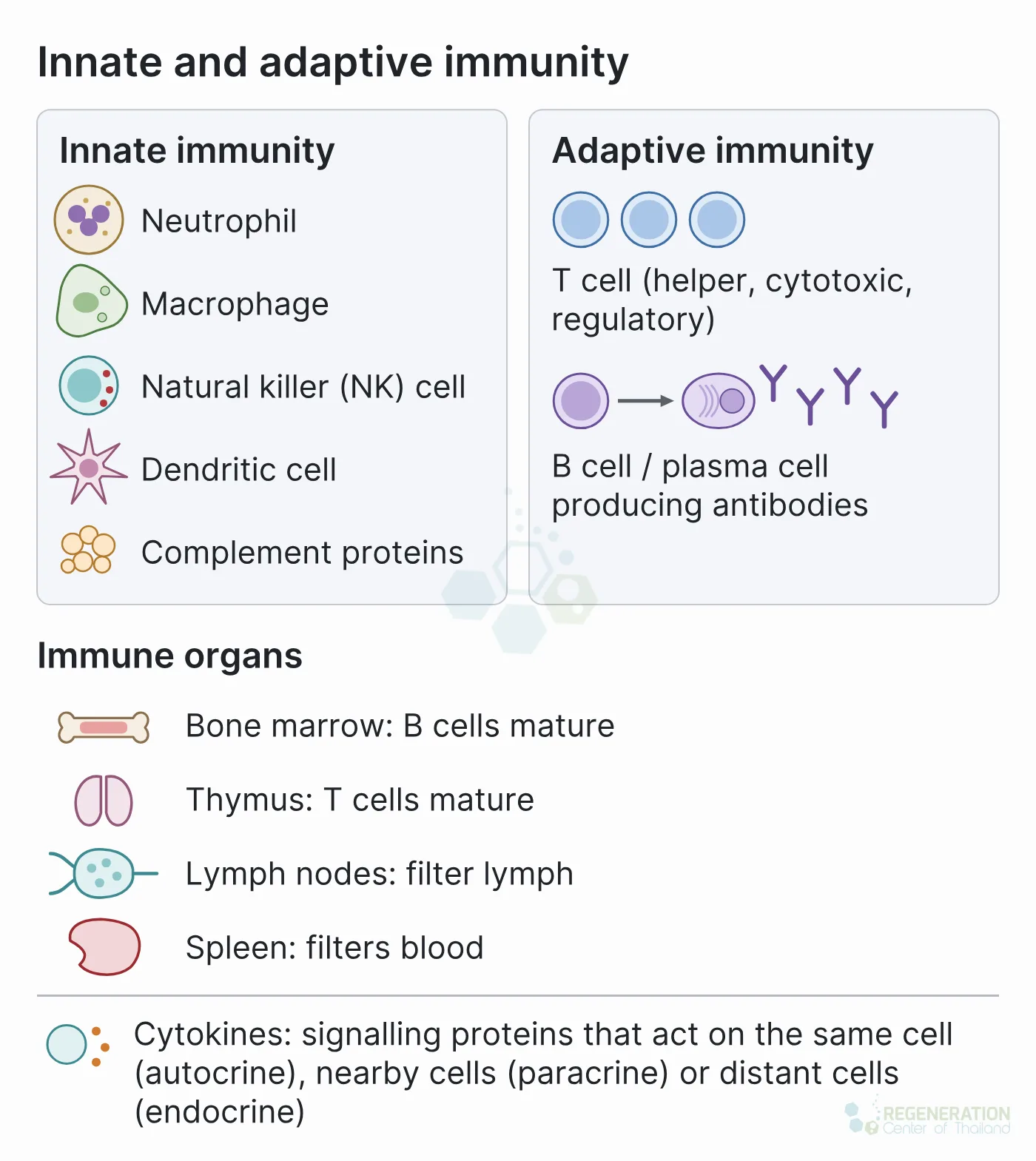
The immune system works through two linked arms. Innate immunity includes cells such as natural killer (NK) cells, while adaptive immunity is coordinated by lymphocytes, mainly B cells and T cells, which recognise specific targets.[6] Both arms distinguish the body’s own tissue from foreign material, and a breakdown in that distinction can injure tissue.[7] B cells mature in the red bone marrow and T cells in the thymus; lymph nodes remove debris and microbes from lymph, and the spleen filters the blood and hosts immune responses against microbes carried in it.[6] After an infection or a vaccine, memory B cells and memory T cells remain, ready to respond faster the next time.[1]
An immune response also has to be stopped. Regulatory T cells (Tregs) are a subset of T cells, made in the thymus or induced from naive T cells elsewhere in the body, that keep the immune system unresponsive to the body’s own antigens and suppress excessive responses. Faulty Treg function is implicated in immune disease.[8] Treatments that modulate immunity either add to these natural controls or override them.
Cytokines: The Signals That Regulate Immunity
Much of this regulation travels through cytokines. The word joins two Greek words, cyto (cell) and kinos (movement). Cytokines are small proteins or glycoproteins released by immune cells and by other cells such as epithelial cells and fibroblasts. There is no single agreed way to classify them; grouped by structure, the main families are the interleukins, the interferons, the tumor necrosis factors (TNF), the colony-stimulating factors, the transforming growth factors and the chemokines, which direct the movement of white blood cells.[9]
A cytokine can act on the cell that released it (autocrine action), on nearby cells (paracrine action) or, in some cases, on distant cells (endocrine action). Different cell types often release the same cytokine, and one cytokine often acts on several cell types.[10] Cytokines are sometimes called the hormones of the immune system, but they differ from classic hormones in two ways. A hormone is mainly the product of one gland or tissue, whereas most cells can make cytokines; and, molecule for molecule, cytokines are far more potent. Interleukin-1, for example, induces the gene for cyclooxygenase-2 (COX-2) at a concentration of about 10 picomolar.[11]
Cytokines and other growth factors also control blood-forming stem cells: whether they stay dormant (quiescence), copy themselves (self-renewal), mature (differentiation), die (apoptosis) or leave their stem cell niche in the marrow (mobilization).[12] One colony-stimulating factor, G-CSF, is given as a medicine to raise neutrophil counts and to mobilize blood stem cells into the bloodstream for collection.[13]
Approved Immunomodulatory Drug Classes
Medicines that change immune activity fall into a few broad groups. The table gives examples; each has its own approved uses, monitoring and risks.
| Direction | Drug class | Examples | How it works |
|---|---|---|---|
| Strengthens | Vaccines | Many, against bacteria and viruses | Expose the immune system to a harmless form or part of a microbe, so antibodies and memory cells are ready before infection[1] |
| Strengthens | Colony-stimulating factors | Filgrastim (G-CSF) | Increases neutrophil production; used to lower the risk of infection during some cancer chemotherapy[13] |
| Strengthens | Immune checkpoint inhibitors | Antibodies that block CTLA-4 or PD-1 | Remove inhibitory signals that limit T cells directed against a tumor[2] |
| Suppresses | Corticosteroids (glucocorticoids) | Prednisolone, methylprednisolone[14] | Bind the glucocorticoid receptor and block the transcription of many cytokines[3] |
| Suppresses | Calcineurin inhibitors | Ciclosporin, tacrolimus | Block the calcineurin signal that switches on T cell genes after activation; central to organ transplantation[3] |
| Suppresses | Antimetabolites | Azathioprine, mycophenolate, methotrexate | Interfere with the DNA synthesis that dividing lymphocytes depend on[3]; methotrexate is the recommended first drug for rheumatoid arthritis, with any glucocorticoid given while it takes effect kept to the lowest dose for the shortest time[15][16] |
| Suppresses | Cytokine blockers | TNF inhibitors (adalimumab, etanercept, infliximab and others); IL-6 receptor blockers (tocilizumab, sarilumab) | Neutralise a single inflammatory cytokine or its receptor; used in rheumatoid arthritis and other inflammatory diseases[15] |
| Suppresses | Co-stimulation blockers | Abatacept, belatacept | Fusion proteins built from CTLA-4, the natural T cell brake, that block the CD80/86 to CD28 co-stimulation signal T cells need for full activation[3] |
| Suppresses | B cell depletion | Rituximab (anti-CD20) | Removes CD20-carrying B cells, usually for up to 6 to 9 months and occasionally longer[3] |
| Suppresses | Janus kinase (JAK) inhibitors | Tofacitinib, baricitinib, upadacitinib | Tablets that block signalling inside cells from several cytokine receptors[15] |
Several of these drugs, and their risks, are discussed condition by condition on our overview of autoimmune diseases. How inflammation itself starts and resolves is covered on our page on inflammation and healing.
The immunomodulatory properties of stem cells
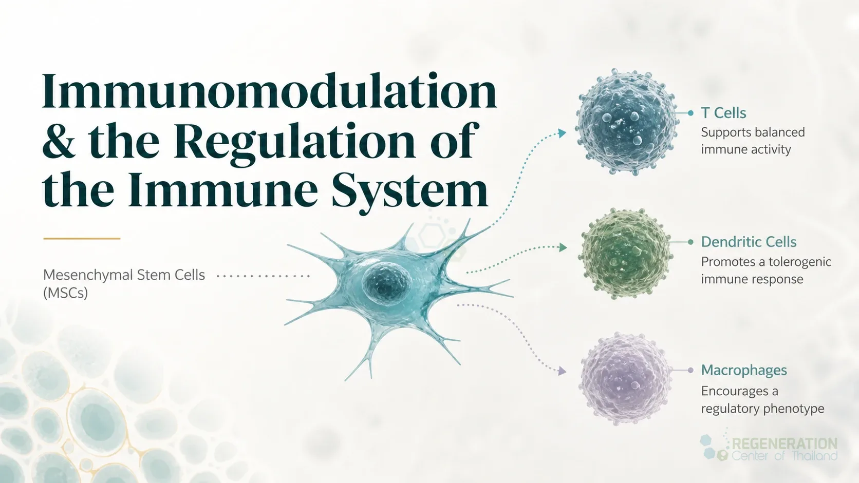
The immune effects of mesenchymal stromal cells are being investigated in inflammatory disorders such as Crohn’s disease and graft-versus-host disease. Their immunomodulatory activity is not fixed: it is switched on, or “licensed”, by inflammatory signals in their surroundings, so the effect of the same cells can differ with the type and level of inflammation in the tissue.[4] In studies of human MSCs grown in the laboratory, suppression of T cells and NK cells required interferon-gamma released by the activated immune cells themselves.[17] In mouse MSCs, interferon-gamma had to be combined with a second inflammatory cytokine, TNF-alpha or interleukin-1, before the cells became suppressive.[18]
The effector molecules also differ between species. Human and monkey MSCs suppressed immune cells mainly through the enzyme indoleamine 2,3-dioxygenase (IDO), while mouse MSCs used nitric oxide under the same culture conditions.[19] This matters because much of the evidence for MSC immunomodulation comes from mice.
Infused MSCs do not stay long in the body. In mice, MSCs injected into a vein were found in the lungs within an hour, and living cells were recovered only from the lungs and only for up to 24 hours; by 24 hours the radioactive label from the cells had moved mainly to the liver, which the authors read as cell debris.[20] In a mouse model of graft-versus-host disease, the cells’ suppressive effect depended on the recipient’s immune cells killing them: phagocytes that engulfed the dying MSCs then produced IDO. In patients with graft-versus-host disease given MSCs, those whose blood cells killed MSCs strongly in a laboratory test were the ones who responded.[21] How the cells relate to the supporting tissue of the marrow and other organs is described on our page on stromal cells and immune regulation.
Mesenchymal stem cells and immunomodulation
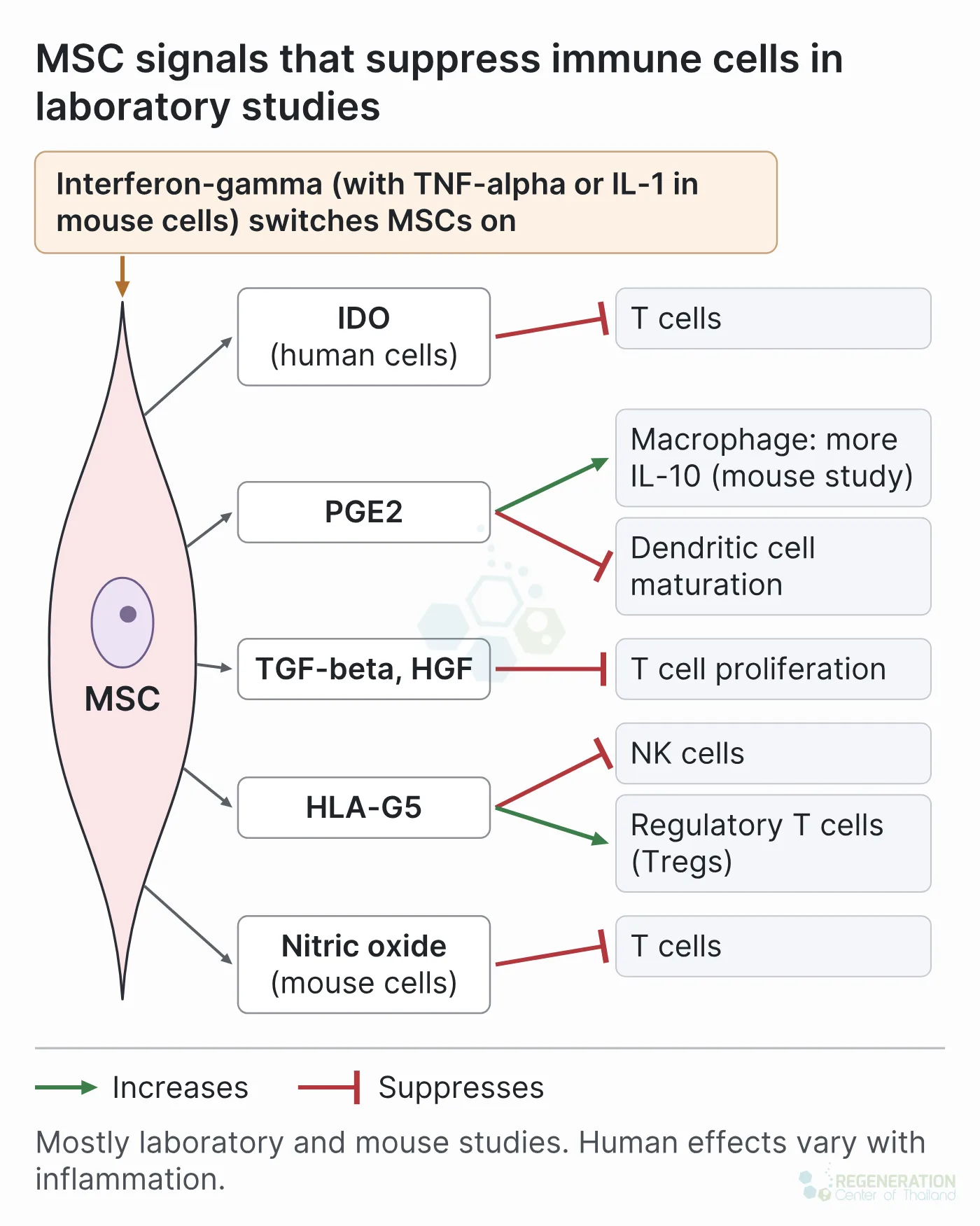
Mesenchymal stem cells, also called mesenchymal stromal cells, act on both arms of the immune system. The best-described mechanisms are listed below with the kind of evidence behind each.
| Mechanism | What was observed | Evidence |
|---|---|---|
| Licensing by interferon-gamma | MSCs became suppressive only after exposure to interferon-gamma from activated T cells and NK cells[17] | Human cells in the laboratory |
| IDO | Interferon-gamma induced IDO, which breaks down tryptophan; adding tryptophan back restored T cell proliferation[22] | Human cells in the laboratory |
| Prostaglandin E2 (PGE2) | MSCs shifted dendritic cells, helper T cells and NK cells toward a less inflammatory profile; blocking PGE2 production reduced the effect[23] | Human cells in the laboratory |
| TGF-beta and hepatocyte growth factor | Suppressed T cell proliferation without contact between the cells; neutralising antibodies identified TGF-beta1 and HGF as the mediators[24] | Human cells in the laboratory |
| HLA-G5 | A soluble HLA-G molecule released by MSCs helped suppress T cells and NK cells[25] | Human cells in the laboratory |
| Regulatory T cell induction | The share of regulatory T cells rose in co-cultures with MSCs[23], and HLA-G5 contributed to the expansion of CD4+CD25high FOXP3+ Tregs[25] | Human cells in the laboratory |
| Shift of macrophages toward repair | In septic mice, MSCs released PGE2 that made the animals’ macrophages produce more of the anti-inflammatory cytokine interleukin-10; survival improved, and the benefit was lost when macrophages or interleukin-10 were removed[26] | Mice |
| Nitric oxide and chemokines | Chemokines drew T cells close to MSCs, where nitric oxide suppressed them; the cells prevented graft-versus-host disease in mice[18] | Mice |
More on macrophage states is on our page on macrophage markers and activation, and on natural killer cells on our page on NK cells. These laboratory and animal results describe possible mechanisms. In advanced clinical trials, MSCs have fallen short of the expectations raised by encouraging animal studies across a wide range of diseases.[27] In the SMART-MS trial, a randomized, double-blind, placebo-controlled study in 18 people with progressive multiple sclerosis, a single injection of the patients’ own bone marrow MSCs into the spinal fluid had no effect on the main measure, the combined evoked-potential latency at 6 months (a test of nerve signal conduction); 7 patients had abnormal spinal MRI findings at 6 months, one developed chronic pain from arachnoiditis, and the authors advised caution with this route.[28]
Human Evidence: Graft-Versus-Host Disease
The clearest human evidence for MSC immunomodulation comes from acute graft-versus-host disease (GVHD), in which immune cells from a donor blood stem cell transplant attack the recipient’s skin, gut and liver.
The largest randomized trial was negative. In a phase 3 trial, 260 patients aged 6 months to 70 years with steroid-refractory acute GVHD were randomized 2 to 1 to the bone marrow MSC product remestemcel-L or placebo, on top of standard second-line treatment. Durable complete response, the main measure, occurred in 35% with cells and 30% with placebo, a difference that was not statistically significant, and overall response at day 28, among the 244 patients who received at least one infusion, was also similar (58% versus 54%). In analyses done after the trial, the 14 children given cells had a higher overall response than the 13 given placebo (64% versus 23%).[29]
Earlier, an open European phase II study had treated 55 patients with steroid-resistant severe acute GVHD; 30 had a complete response and 9 improved.[30] A single-arm phase 3 study then gave remestemcel-L to 54 children whose acute GVHD had not responded to steroids. By day 28, 70.4% had responded, against a pre-set benchmark of 45%; there was no placebo group.[31]
On 18 December 2024 the US Food and Drug Administration approved remestemcel-L (Ryoncil) for steroid-refractory acute GVHD in children aged 2 months and older, on the basis of that 54-child study (30% complete and 41% partial responses at day 28). The most common side effects were infections, fever, bleeding, swelling, abdominal pain and high blood pressure.[32] Remestemcel-L is on the FDA list of approved cellular and gene therapy products, current as of 30 October 2026, and is the first and so far only mesenchymal stromal cell therapy the FDA has approved; its approval does not extend to adults or to any autoimmune disease.[32][5] In Japan, a bone marrow MSC product (Temcell) is approved for acute GVHD; of 306 evaluable patients in a Japanese cellular therapy registry, 56% had responded by day 28, and 40% of all 381 treated patients were alive at 6 months.[33]
Outside GVHD, one MSC product, darvadstrocel (donor fat-derived MSCs injected into Crohn’s disease perianal fistulas), held a European marketing authorisation. Its confirmatory phase 3 trial in 568 patients found no benefit: combined remission at 24 weeks in 48.8% with cells and 46.3% with placebo.[34] Its European marketing authorisation was withdrawn, at the company’s request, on 13 December 2024.[35] Trials of MSCs in rheumatoid arthritis, lupus, multiple sclerosis and other autoimmune diseases are summarised, condition by condition, on our autoimmune disease overview.
Published Clinical Citations
[1] ^ Pollard AJ, Bijker EM. A guide to vaccinology: from basic principles to new developments. Nature Reviews Immunology. 2021;21(2):83-100. https://doi.org/10.1038/s41577-020-00479-7
[2] ^ Ribas A, Wolchok JD. Cancer immunotherapy using checkpoint blockade. Science. 2018;359(6382):1350-1355. https://doi.org/10.1126/science.aar4060
[3] ^ Wiseman AC. Immunosuppressive medications. Clinical Journal of the American Society of Nephrology. 2016;11(2):332-343. https://doi.org/10.2215/CJN.08570814
[4] ^ Shi Y, Wang Y, Li Q, et al. Immunoregulatory mechanisms of mesenchymal stem and stromal cells in inflammatory diseases. Nature Reviews Nephrology. 2018;14(8):493-507. https://doi.org/10.1038/s41581-018-0023-5
[5] ^ US Food and Drug Administration. Approved Cellular and Gene Therapy Products. Content current as of 30 October 2026. https://www.fda.gov/vaccines-blood-biologics/cellular-gene-therapy-products/approved-cellular-and-gene-therapy-products
[6] ^ Betts JG, Young KA, Wise JA, et al. Anatomy and Physiology 2e. Section 21.1, Anatomy of the Lymphatic and Immune Systems. OpenStax, Rice University. https://openstax.org/books/anatomy-and-physiology-2e/pages/21-1-anatomy-of-the-lymphatic-and-immune-systems
[7] ^ Chaplin DD. Overview of the immune response. Journal of Allergy and Clinical Immunology. 2010;125(2 Suppl 2):S3-S23. https://doi.org/10.1016/j.jaci.2009.12.980
[8] ^ Sakaguchi S, Yamaguchi T, Nomura T, Ono M. Regulatory T cells and immune tolerance. Cell. 2008;133(5):775-787. https://doi.org/10.1016/j.cell.2008.05.009
[9] ^ Gonçalves Dos Santos G, Delay L, Yaksh TL, Corr M. Neuraxial cytokines in pain states. Frontiers in Immunology. 2019;10:3061. https://doi.org/10.3389/fimmu.2019.03061
[10] ^ Zhang JM, An J. Cytokines, inflammation, and pain. International Anesthesiology Clinics. 2007;45(2):27-37. https://doi.org/10.1097/AIA.0b013e318034194e
[11] ^ Dinarello CA. Historical insights into cytokines. European Journal of Immunology. 2007;37(Suppl 1):S34-S45. https://doi.org/10.1002/eji.200737772
[12] ^ Zhang CC, Lodish HF. Cytokines regulating hematopoietic stem cell function. Current Opinion in Hematology. 2008;15(4):307-311. https://doi.org/10.1097/MOH.0b013e3283007db5
[13] ^ Amgen Inc. NEUPOGEN (filgrastim) injection, for subcutaneous or intravenous use. US prescribing information, revised July 2026. DailyMed set ID 97cc73cc-b5b7-458a-a933-77b00523e193. https://dailymed.nlm.nih.gov/dailymed/drugInfo.cfm?setid=97cc73cc-b5b7-458a-a933-77b00523e193
[14] ^ Deng D, Zhang P, Guo Y, Lim TO. A randomised double-blind, placebo-controlled trial of allogeneic umbilical cord-derived mesenchymal stem cell for lupus nephritis. Annals of the Rheumatic Diseases. 2017;76(8):1436-1439. https://doi.org/10.1136/annrheumdis-2017-211073
[15] ^ Smolen JS, Landewé RBM, Bergstra SA, et al. EULAR recommendations for the management of rheumatoid arthritis with synthetic and biological disease-modifying antirheumatic drugs: 2022 update. Annals of the Rheumatic Diseases. 2023;82(1):3-18. https://doi.org/10.1136/ard-2022-223356
[16] ^ Fraenkel L, Bathon JM, England BR, et al. 2021 American College of Rheumatology guideline for the treatment of rheumatoid arthritis. Arthritis Care & Research. 2021;73(7):924-939. https://doi.org/10.1002/acr.24596
[17] ^ Krampera M, Cosmi L, Angeli R, et al. Role for interferon-gamma in the immunomodulatory activity of human bone marrow mesenchymal stem cells. Stem Cells. 2006;24(2):386-398. https://doi.org/10.1634/stemcells.2005-0008
[18] ^ Ren G, Zhang L, Zhao X, et al. Mesenchymal stem cell-mediated immunosuppression occurs via concerted action of chemokines and nitric oxide. Cell Stem Cell. 2008;2(2):141-150. https://doi.org/10.1016/j.stem.2007.11.014
[19] ^ Ren G, Su J, Zhang L, et al. Species variation in the mechanisms of mesenchymal stem cell-mediated immunosuppression. Stem Cells. 2009;27(8):1954-1962. https://doi.org/10.1002/stem.118
[20] ^ Eggenhofer E, Benseler V, Kroemer A, et al. Mesenchymal stem cells are short-lived and do not migrate beyond the lungs after intravenous infusion. Frontiers in Immunology. 2012;3:297. https://doi.org/10.3389/fimmu.2012.00297
[21] ^ Galleu A, Riffo-Vasquez Y, Trento C, et al. Apoptosis in mesenchymal stromal cells induces in vivo recipient-mediated immunomodulation. Science Translational Medicine. 2017;9(416):eaam7828. https://doi.org/10.1126/scitranslmed.aam7828
[22] ^ Meisel R, Zibert A, Laryea M, et al. Human bone marrow stromal cells inhibit allogeneic T-cell responses by indoleamine 2,3-dioxygenase-mediated tryptophan degradation. Blood. 2004;103(12):4619-4621. https://doi.org/10.1182/blood-2003-11-3909
[23] ^ Aggarwal S, Pittenger MF. Human mesenchymal stem cells modulate allogeneic immune cell responses. Blood. 2005;105(4):1815-1822. https://doi.org/10.1182/blood-2004-04-1559
[24] ^ Di Nicola M, Carlo-Stella C, Magni M, et al. Human bone marrow stromal cells suppress T-lymphocyte proliferation induced by cellular or nonspecific mitogenic stimuli. Blood. 2002;99(10):3838-3843. https://doi.org/10.1182/blood.v99.10.3838
[25] ^ Selmani Z, Naji A, Zidi I, et al. Human leukocyte antigen-G5 secretion by human mesenchymal stem cells is required to suppress T lymphocyte and natural killer function and to induce CD4+CD25highFOXP3+ regulatory T cells. Stem Cells. 2008;26(1):212-222. https://doi.org/10.1634/stemcells.2007-0554
[26] ^ Németh K, Leelahavanichkul A, Yuen PS, et al. Bone marrow stromal cells attenuate sepsis via prostaglandin E(2)-dependent reprogramming of host macrophages to increase their interleukin-10 production. Nature Medicine. 2009;15(1):42-49. https://doi.org/10.1038/nm.1905
[27] ^ Galipeau J, Sensébé L. Mesenchymal stromal cells: clinical challenges and therapeutic opportunities. Cell Stem Cell. 2018;22(6):824-833. https://doi.org/10.1016/j.stem.2018.05.004
[28] ^ Kvistad CE, Kråkenes T, Holmøy T, et al. Intrathecal mesenchymal stem cells in progressive multiple sclerosis: a randomized, double-blind, placebo-controlled trial (SMART-MS). Neurology. 2026;106(10):e214915. https://doi.org/10.1212/WNL.0000000000214915
[29] ^ Kebriaei P, Hayes J, Daly A, et al. A phase 3 randomized study of remestemcel-L versus placebo added to second-line therapy in patients with steroid-refractory acute graft-versus-host disease. Biology of Blood and Marrow Transplantation. 2020;26(5):835-844. https://doi.org/10.1016/j.bbmt.2019.08.029
[30] ^ Le Blanc K, Frassoni F, Ball L, et al. Mesenchymal stem cells for treatment of steroid-resistant, severe, acute graft-versus-host disease: a phase II study. Lancet. 2008;371(9624):1579-1586. https://doi.org/10.1016/S0140-6736(08)60690-X
[31] ^ Kurtzberg J, Abdel-Azim H, Carpenter P, et al. A phase 3, single-arm, prospective study of remestemcel-L, ex vivo culture-expanded adult human mesenchymal stromal cells for the treatment of pediatric patients who failed to respond to steroid treatment for acute graft-versus-host disease. Biology of Blood and Marrow Transplantation. 2020;26(5):845-854. https://doi.org/10.1016/j.bbmt.2020.01.018
[32] ^ US Food and Drug Administration. FDA approves first mesenchymal stromal cell therapy to treat steroid-refractory acute graft-versus-host disease. News release, 18 December 2024. https://www.fda.gov/news-events/press-announcements/fda-approves-first-mesenchymal-stromal-cell-therapy-treat-steroid-refractory-acute-graft-versus-host
[33] ^ Murata M, Terakura S, Wake A, et al. Off-the-shelf bone marrow-derived mesenchymal stem cell treatment for acute graft-versus-host disease: real-world evidence. Bone Marrow Transplantation. 2021;56(10):2355-2366. https://doi.org/10.1038/s41409-021-01304-y
[34] ^ Colombel JF, Garcia Olmo D, Chen ST, et al. Darvadstrocel in patients with Crohn's disease with complex perianal fistulas: the ADMIRE CD II phase 3 randomized trial. Gastroenterology. 2026;170(7):1562-1570. https://doi.org/10.1053/j.gastro.2025.12.033
[35] ^ European Medicines Agency. Alofisel (darvadstrocel): EPAR. Withdrawal of the marketing authorisation in the European Union, 13 December 2024. https://www.ema.europa.eu/en/medicines/human/EPAR/alofisel