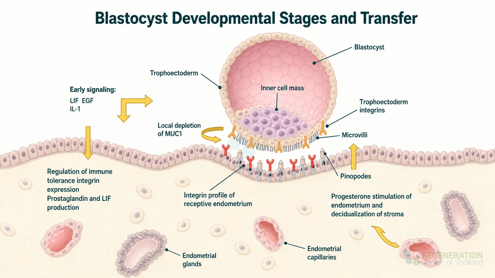
A blastocyst is an embryo about five to seven days after fertilization. It is a hollow ball of cells with three parts: an outer layer of trophectoderm cells that will form the trophoblast, the main tissue of the placenta, a small cluster of cells inside, the inner cell mass, that will form the embryo itself, and a fluid-filled cavity, the blastocoel. In humans it forms on day 5, hatches from its protective shell on day 6 and implants in the wall of the uterus around day 7.[1] In IVF, embryos are often grown to this stage in the laboratory before one is selected for transfer, and the inner cell mass of the blastocyst is the source of human embryonic stem cells.
Structure of the Blastocyst
The blastocyst is the first stage at which the embryo’s different cell types can be told apart under the microscope.
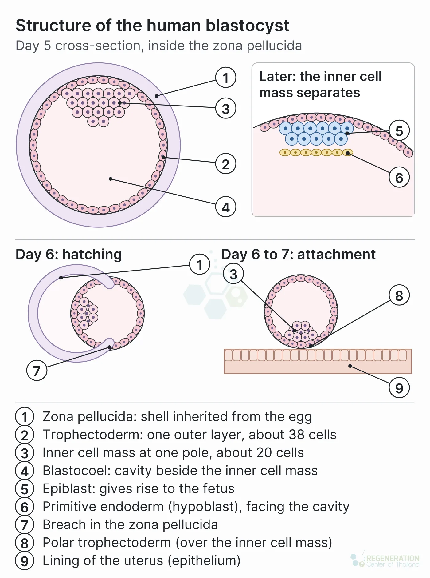
- Trophectoderm: the single outer layer of cells. These are extraembryonic cells: after implantation they give rise to the placental trophoblast, not to the fetus.[1] The trophoblast forms most of the placenta; in primates, extraembryonic mesoderm from the inner cell mass lineages also becomes part of the chorion and allantois, the fetal side of the placenta.[2] How they go on to form the placenta is described on our page on trophoblast differentiation.
- Inner cell mass: a cluster of pluripotent cells attached to the inside of the trophectoderm at one pole. By the time the blastocyst implants, it has separated into the epiblast, which gives rise to all the tissues of the fetus, and the primitive endoderm (hypoblast), an extraembryonic layer that does not contribute to the fetus.[1]
- Blastocoel: the fluid-filled cavity that opens up inside the embryo as the blastocyst forms and then expands.
- Zona pellucida: the glycoprotein shell inherited from the egg, which still surrounds the blastocyst until hatching.
A human blastocyst is small in cell number. In a laboratory study of surplus IVF embryos, newly expanded blastocysts had on average 58 cells on day 5, 84 on day 6 and 125 on day 7. The trophectoderm had about 38 cells on day 5 and about 81 on day 7, while the inner cell mass grew from about 20 to about 42 cells between days 5 and 6. Cell death was common in both parts.[3]
From Morula to Blastocyst
The embryo arrives at this stage through a fixed sequence, described step by step on our page on zygote formation. Three cleavage divisions take the fertilized egg from one cell to eight by day 3, when the embryo’s own genes switch on. On or around day 4 the cells compact into a tight ball, the morula. On day 5 fluid collects inside it, and the outer and inner cells become trophectoderm and inner cell mass.[1]
Many human embryos never get this far. In IVF culture, 50 to 70 percent of embryos routinely fail to reach the blastocyst stage.[1] In the cell-counting study above, 63 of 149 normally fertilized embryos (42 percent) reached the blastocyst stage by day 5 or 6.[3]
Hatching and Implantation
On day 6 the expanding blastocyst breaks out of the zona pellucida, a step called hatching, and it is ready to implant on day 7.[1] In humans the trophectoderm over the inner cell mass (the polar trophectoderm) attaches to the lining of the uterus at about 6 to 7 days after fertilization. Its cells fuse into a primary syncytium, a layer of merged cells with many nuclei, which quickly invades through the surface lining into the tissue beneath.[4] Mice differ: in the mouse it is the trophectoderm on the opposite side, away from the inner cell mass, that attaches first.[4] Around the same time the inner cell mass begins to reorganize into the two-layered disc from which the embryo develops; that next stage, and the formation of the three germ layers, is described on our page on germ layers and gastrulation.
Blastocyst Grading in IVF
Embryologists rank blastocysts by their appearance under the microscope, using a three-part system first described by Gardner and Schoolcraft. It scores how far the blastocyst has expanded, then grades the inner cell mass and the trophectoderm separately. Gardner’s letters A, B and C were turned into the numbers 1, 2 and 3 in an international consensus of embryologists in 2011, the Istanbul consensus, which also set day 5 assessment at 116 hours (plus or minus 2) after insemination.[5] A Gardner-style grade such as “4AA” combines a number for expansion with the two letters, so “AA” means a good inner cell mass and a good trophectoderm.
| Feature graded | Good (Gardner A, consensus 1) | Fair (B, 2) | Poor (C, 3) |
|---|---|---|---|
| Inner cell mass | Prominent and easily seen, many cells compacted and tightly adhered | Easily seen, many cells loosely grouped | Difficult to see, few cells |
| Trophectoderm | Many cells forming a cohesive epithelium | Few cells forming a loose epithelium | Very few cells |
Expansion is recorded on a separate scale; the consensus groups the stages as early, blastocyst, expanded, and hatching or hatched.[5] In a 2025 update, the same groups revised these recommendations to cover time-lapse imaging and advised that several less-studied features either not be used for grading or be used only to rank embryos rather than to grade them.[6]
Grade is linked to the chance of implantation, but it is a prediction, not a guarantee. In Gardner’s own retrospective series of 107 patients who each had two blastocysts transferred, implantation rates were 70 percent per embryo when both were top-scoring, 50 percent when one was, and 28 percent when only low-scoring blastocysts were available; twin pregnancies occurred in 61, 50 and 29 percent of those groups. The authors concluded that transferring one high-scoring blastocyst should give pregnancy rates above 60 percent without the complication of twins.[7]
Blastocyst Transfer: What the Evidence Shows
Blastocyst transfer means culturing embryos for five or six days and placing one in the uterus at the blastocyst stage, rather than on day 2 or 3 at the cleavage stage. The idea is that longer culture lets embryos that cannot develop further stop on their own, and that a blastocyst better matches the timing of the uterine lining.[8]
A Cochrane systematic review of 32 randomized trials (5,821 women or couples), with searches to October 2021, found:[8]
- Live birth after a fresh transfer: higher with blastocyst transfer (odds ratio 1.27; 15 trials, 2,219 women). If 31 percent of women have a live birth after a fresh cleavage-stage transfer, 32 to 41 percent would after a blastocyst transfer. The evidence was rated low quality.
- Cumulative pregnancy from one egg collection, counting later frozen transfers: uncertain whether the stage of transfer makes a difference.
- No embryo to transfer: more frequent with blastocyst culture, rising from about 1 percent to between 2 and 4 percent of women.
- Embryos left to freeze: fewer after blastocyst culture (from 60 percent of women to between 37 and 46 percent).
- Multiple pregnancy and miscarriage: uncertain overall; in the lower-risk-of-bias trials transferring equal numbers of embryos, blastocyst transfer probably increased multiple pregnancy.
On the number of embryos to transfer, the European Society of Human Reproduction and Embryology (ESHRE) guideline of 2024 found that no clinical or embryological factor by itself justifies transferring two embryos instead of one, because double transfer raises the rate of multiple pregnancy and with it preterm birth and low birthweight. It recommends a single embryo for a frozen-thawed blastocyst transfer regardless of the blastocyst’s quality, and states that morphology should not be the reason for transferring two fresh blastocysts.[9] Results for any individual depend on age, the number and quality of embryos and the clinic, and are a matter for a fertility specialist.
The Inner Cell Mass and Embryonic Stem Cells
Human embryonic stem cells are usually derived by culturing the inner cell mass of a blastocyst in the laboratory, and most attempts fail at the first steps.[1] The first human lines derived from blastocysts were reported in 1998. They kept normal chromosomes and, after 4 to 5 months of growth in culture, could still form trophoblast and derivatives of all three germ layers: gut lining (endoderm), cartilage, bone and muscle (mesoderm), and nerve tissue and skin-type epithelium (ectoderm).[10] The inner cell mass itself is pluripotent: it forms every tissue of the fetus but not the trophoblast, which comes from the trophectoderm.[1] In culture, human embryonic stem cells can also make some trophoblast cells.[10]
Published Clinical Citations
[1] ^ Niakan KK, Han J, Pedersen RA, Simon C, Reijo Pera RA. Human pre-implantation embryo development. Development. 2012;139(5):829-841. https://doi.org/10.1242/dev.060426
[2] ^ Ross C, Boroviak TE. Origin and function of the yolk sac in primate embryogenesis. Nature Communications. 2020;11(1):3760. https://doi.org/10.1038/s41467-020-17575-w
[3] ^ Hardy K, Handyside AH, Winston RM. The human blastocyst: cell number, death and allocation during late preimplantation development in vitro. Development. 1989;107(3):597-604. https://doi.org/10.1242/dev.107.3.597
[4] ^ Turco MY, Moffett A. Development of the human placenta. Development. 2019;146(22):dev163428. https://doi.org/10.1242/dev.163428
[5] ^ Alpha Scientists in Reproductive Medicine and ESHRE Special Interest Group of Embryology. The Istanbul consensus workshop on embryo assessment: proceedings of an expert meeting. Human Reproduction. 2011;26(6):1270-1283. https://doi.org/10.1093/humrep/der037
[6] ^ ESHRE Special Interest Group of Embryology and Alpha Scientists in Reproductive Medicine, Coticchio G, Ahlström A, et al. The Istanbul Consensus update: a revised ESHRE/ALPHA consensus on oocyte and embryo static and dynamic morphological assessment. Reproductive BioMedicine Online. 2025;50(6):104955. https://doi.org/10.1016/j.rbmo.2025.104955
[7] ^ Gardner DK, Lane M, Stevens J, Schlenker T, Schoolcraft WB. Blastocyst score affects implantation and pregnancy outcome: towards a single blastocyst transfer. Fertility and Sterility. 2000;73(6):1155-1158. https://doi.org/10.1016/s0015-0282(00)00518-5
[8] ^ Glujovsky D, Quinteiro Retamar AM, Alvarez Sedo CR, Ciapponi A, Cornelisse S, Blake D. Cleavage-stage versus blastocyst-stage embryo transfer in assisted reproductive technology. Cochrane Database of Systematic Reviews. 2022;5(5):CD002118. https://doi.org/10.1002/14651858.CD002118.pub6
[9] ^ ESHRE Guideline Group on the Number of Embryos to Transfer, Alteri A, Arroyo G, et al. ESHRE guideline: number of embryos to transfer during IVF/ICSI. Human Reproduction. 2024;39(4):647-657. https://doi.org/10.1093/humrep/deae010
[10] ^ Thomson JA, Itskovitz-Eldor J, Shapiro SS, et al. Embryonic stem cell lines derived from human blastocysts. Science. 1998;282(5391):1145-1147. https://doi.org/10.1126/science.282.5391.1145