
Oligodendrocytes are particular useful types of cell for treatment in conditions such as ataxia,brain injuries,ALS,motor neuron disease and spinal cord damage.
[1] Oligodendrocytes are also helpful for neural function in that it provides insulation through the myelin sheath.
For instance, in our treatments of autoimmune diseases like multiple sclerosis, considerable damage in the myelin sheath may be damaged, which exposes the neural cell underneath and impairs nerve signal transmission. [2]
Myelin Sheath Repair Video
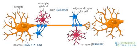
Oligodendrocytes are the myelin-producing cells of the central nervous system (CNS). These cells play a critical role in neural signal transmission and overall CNS function. Myelination by oligodendrocytes increases the speed at which electrical signals travel along neurons, allowing for efficient neural communication. Differentiation of oligodendrocyte progenitor cells (OPCs) into mature oligodendrocytes and subsequent myelination is a complex process that is regulated by a combination of intrinsic and extrinsic factors. This process can be tracked and studied using various markers at different stages of differentiation.
- Oligodendrocyte Progenitor Markers: PDGFRα (Platelet Derived Growth Factor Receptor Alpha): A receptor for PDGF, which is a growth factor that stimulates the proliferation of OPCs.
- NG2 (Neuron-glial antigen 2): A proteoglycan found on the surface of OPCs.
- Immature/Mature Oligodendrocyte Markers: O4: This antigen appears early in oligodendrocyte differentiation, even before the cells begin to myelinate axons.
- GalC (Galactocerebroside): Appears slightly later than O4 and is considered a marker of differentiation.
- MBP (Myelin Basic Protein): A major component of the myelin sheath, MBP is expressed as oligodendrocytes begin the myelination process.
- Myelin Markers:MOG (Myelin Oligodendrocyte Glycoprotein): A glycoprotein believed to be important for the stability of the myelin sheath.
- PLP (Proteolipid Protein): Another major myelin protein, with essential roles in myelin function.
Mechanisms of Myelination
After differentiation, oligodendrocytes start the process of myelination, where they extend processes that wrap around the axons of neurons. These wraps consist of multiple layers of membrane rich in lipids, forming the myelin sheath. This myelin sheath acts as an insulator, which facilitates the rapid conduction of electrical impulses along the axon.
Several factors influence the myelination process:
- Neuronal Signals: Neurons and axons release signals that can promote or inhibit myelination.
- Extrinsic Factors: These include cytokines, growth factors, and other signaling molecules that can either promote or inhibit the differentiation and myelination processes.
- Transcription Factors: Factors such as Olig1, Olig2, and Nkx2.2 play essential roles in oligodendrocyte differentiation.
- Pathology: Any disturbances in the differentiation of OPCs into mature oligodendrocytes or in the myelination process can lead to several CNS disorders, such as multiple sclerosis, where the immune system attacks myelin sheaths.
Research in oligodendrocyte biology aims to understand the mechanisms underlying differentiation and myelination and to identify therapeutic targets for demyelinating diseases.
Published Clinical Citations
[1] ^ Mosebach, Jennifer, Gerburg Keilhoff, Tomasz Gos, Kolja Schiltz, Linda Schoeneck, Henrik Dobrowolny, Christian Mawrin, et al. 2013. Increased nuclear Olig1-expression in the pregenual anterior cingulate white matter of patients with major depression: a regenerative attempt to compensate oligodendrocyte loss? Journal of psychiatric research, no. 8 (April 21). doi:10.1016/j.jpsychires.2013.03.018. https://www.ncbi.nlm.nih.gov/pubmed/23615187
[2] ^ Pérez, María Julia, Natalia Fernandez, and Juana María Pasquini. 2013. Oligodendrocyte differentiation and signaling after transferrin internalization: a mechanism of action. Experimental neurology (June 21). doi:10.1016/j.expneurol.2013.06.014. https://www.ncbi.nlm.nih.gov/pubmed/23797152