
Please complete the inquiry form below. One of our staff will contact you within 1 business day
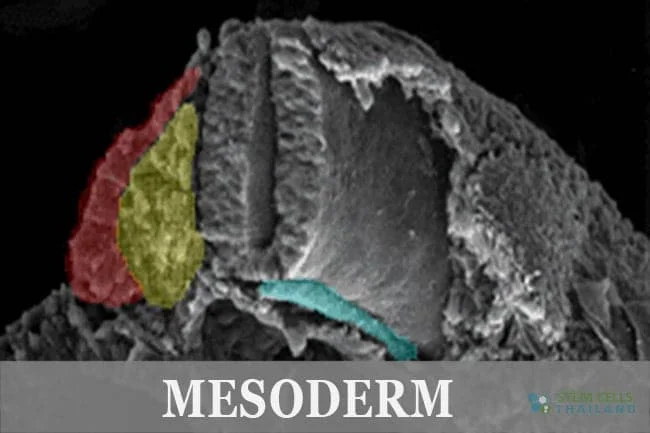
Mesoderm is the The middle part of the three germ layers and a derivative of a blastocyst inner cell mass. The mesodermal layer is composed of cells that have the ability to differentiate into muscle and bone cells, as well as kidney cells ,ligaments and connective tissues.[1]
Mesoderm specifically gives rise to the following structures and tissues:
During embryonic development, the mesoderm originates from the epiblast. Through a process called gastrulation, cells migrate inwards to form this middle germ layer. As development progresses, the mesoderm further differentiates and specializes to form the above-mentioned structures and tissues.
The proper formation and differentiation of the mesoderm are crucial for the development of a healthy embryo. Any disruptions or abnormalities in this process can lead to congenital malformations or developmental disorders.
For humans, the mesoderm is among the 3 primary germ cell layers. The other two germ layers are endoderm and the ectoderm. In the early stages of an embryo the mesoderm area is the central layer.The Mesoderm layer forms during gastrulation and lies between the ectoderm and the endoderm.[2]
It leads to development of some glands and also helps give rise to several other vital structures and tissues including cartilage,bone,connective tissue, muscle, blood for vascular system, reproductive,urinogenital and excretory systems. Stem cells in mesodermal tissues keep the ability to differentiate in varied ways. For instance, Bone marrow stem cells “mesoderm” can become liver cells “endoderm”.[3]
The mesoderm can be identified with 3 germ layers seen in the embryo of all “Bilaterian” creatures. That includes all creatures on Earth with the exception Cnidarians,Placozoans and sponges making them triploblastic. The NODAL indicator helps to mediate the initial formation of the mesoderm layer.
[1] ^ Phermthai, Tatsanee, Singpetch Suksompong, Nednapis Tirawanchai, Surapol Issaragrisil, Suphakde Julavijitphong, Suparat Wichitwiengrat, Decha Silpsorn, and Puttachart Pokathikorn. 2013. Epigenetic analysis and suitability of amniotic fluid stem cells for research and therapeutic purposes. Stem cells and development, no. 9 (February 12). doi:10.1089/scd.2012.0371. https://www.ncbi.nlm.nih.gov/pubmed/23249260
[2] ^ Noisa, Parinya, and Rangsun Parnpai. 2011. Technical challenges in the derivation of human pluripotent cells. Stem cells international (June 19). doi:10.4061/2011/907961. https://www.ncbi.nlm.nih.gov/pubmed/21776284
[3] ^ Moroz, Leonid L, Kevin M Kocot, Mathew R Citarella, Sohn Dosung, Tigran P Norekian, Inna S Povolotskaya, Anastasia P Grigorenko, et al. 2014. The ctenophore genome and the evolutionary origins of neural systems. Nature, no. 7503 (May 21). doi:10.1038/nature13400. https://www.ncbi.nlm.nih.gov/pubmed/24847885
Your liver rarely complains. It filters your blood, breaks down medications, stores energy, and clears… Read More
If you've seen people take ice baths or cold showers and wondered if they're onto… Read More
Immunomodulation stands at the forefront of biomedical research, steering the immune system's ability to fight… Read More
Stem cell research leads the charge in medical innovation, heralding revolutionary advances in regenerative medicine.… Read More
The blood-brain barrier (BBB) is a crucial shield for the brain, regulating the entry of… Read More
While peptide bonds are fundamental to protein structure, their direct relationship with stem cells lies… Read More