


Please complete the inquiry form below. One of our staff will contact you within 1 business day
Dilated cardiomyopathy (DCM) is a serious condition of the heart muscle characterized by the enlargement and dilation of one or both ventricles, along with impaired contractility and reduced systolic function. It is the third most common cause of heart failure and remains the most frequent underlying reason for heart transplantation worldwide, with an estimated prevalence of 40 in every 100,000 persons.
Dilated cardiomyopathy occurs when the heart’s ventricles stretch and thin, becoming enlarged and weakened to the point where the heart can no longer pump blood efficiently throughout the body. Patients with DCM typically have a left ventricular ejection fraction (LVEF) of less than 40%, significantly below the healthy range of 52%–68%, resulting in chronic fatigue, shortness of breath, fluid retention, and markedly diminished quality of life. As the heart chambers dilate, the muscular walls lose their ability to contract with adequate force, resulting in progressively worsening systolic dysfunction. This pathological remodeling process, characterized by gradual increases in left ventricular volume and continuous thinning of the ventricular walls, is typically associated with a steady decline in ejection fraction over time.
Over the past two decades, regenerative stem cell therapy has emerged as a promising alternative approach for patients diagnosed with dilated cardiomyopathy. Published research has demonstrated that mesenchymal stem cells can differentiate into functional cardiomyocytes, promote angiogenesis, reduce myocardial fibrosis, and modulate inflammatory processes that contribute to progressive cardiac deterioration. The Regeneration Center’s UC-MSC+ cardiopoietic cell therapy protocol for DCM aims to restore cardiac function at the cellular level, offering a safe, non-surgical treatment option for patients seeking to improve ventricular performance without the risks associated with open-heart surgery or organ transplantation.
The causes of dilated cardiomyopathy are diverse and can be broadly categorized into ischemic and non-ischemic etiologies. While ischemic DCM results from coronary artery disease, reducing blood flow to the heart muscle, non-ischemic forms account for approximately 41% of all cases and often carry a genetic component. The primary types of dilated cardiomyopathy include:
Idiopathic DCM is the most commonly diagnosed form, occurring in cases where no identifiable secondary cause can be established. Over the past 35 years, however, many cases previously classified as idiopathic have been recognized to have a genetic etiology, with mutations in definitive causal genes identified in up to 40% of these patients. Truncating variants in the TTN gene (encoding the protein titin) are the single most common genetic cause, accounting for approximately 20%–25% of all genetic DCM cases and 18% of sporadic cases.[2]
Familial DCM is defined by the presence of two or more blood relatives diagnosed with the condition, or one relative with DCM combined with a family history of sudden cardiac death before age 35. Approximately 25%–35% of individuals with dilated cardiomyopathy have familial forms of the disease. The inheritance pattern is most commonly autosomal dominant, though variants in more than 20 different genes, many of which encode cytoskeletal and sarcomeric proteins essential for cardiac muscle contraction, have been implicated. The Thailand Regeneration Center offers comprehensive genetic testing and DNA panels for heritable cardiac conditions to help identify familial cardiomyopathy variants.[1]
Peripartum cardiomyopathy (PPCM) represents a distinct form of DCM that develops during the final month of pregnancy or within five months following delivery. Research has demonstrated a significant genetic overlap between PPCM and idiopathic DCM, particularly through shared TTN truncating variants found in approximately 10% of all peripartum cases. This condition is reversible in roughly half of affected patients, though others may progress to chronic dilated cardiomyopathy requiring ongoing management.
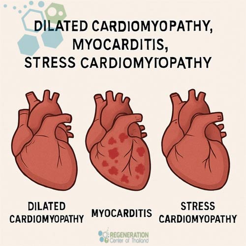
Certain viral infections, including those causing myocarditis, can directly damage cardiomyocytes and trigger an inflammatory cascade that leads to ventricular dilation. When the viral infection fails to clear from cardiac tissue, chronic inflammation and immune-mediated damage can result in progressive DCM. Autoimmune-related cardiomyopathy, in which the immune system mistakenly attacks healthy heart cells, represents another inflammatory pathway to dilated cardiomyopathy.
Exposure to cardiotoxic substances, including chronic excessive alcohol consumption, cocaine use, and certain chemotherapy drugs such as doxorubicin, can induce cardiomyocyte injury and progressive ventricular dilation. Alcoholic cardiomyopathy, in particular, is a well-established form of toxic DCM that may be partially reversible with cessation of alcohol intake and appropriate medical management.
Many patients with early-stage dilated cardiomyopathy remain asymptomatic, making early detection and diagnosis challenging. As the condition progresses and ventricular function continues to decline, patients typically develop symptoms consistent with congestive heart failure, including:
Without timely intervention, dilated cardiomyopathy can lead to several serious complications, including progressive heart failure, cardiac arrhythmias (including life-threatening ventricular tachycardia), heart valve regurgitation caused by chamber dilation distorting valve structures, thromboembolic events (blood clots forming in the dilated chambers that can cause ischemic or hemorrhagic stroke), and sudden cardiac arrest. The overall prognosis for patients with DCM remains guarded — approximately 50% of patients with advanced disease face mortality within five years, and many ultimately become candidates for heart transplantation or mechanical assist devices.
Dilated cardiomyopathy and ischemic heart disease can impact numerous organs in the body. The injured areas of the heart directly affected by DCM cannot produce enough blood for the kidneys, which then affects their ability to excrete water and salt (sodium). This disturbed kidney function may cause patients to retain excess fluid. Patients with higher cardiometabolic risk factors may also develop pulmonary edema (PE), which occurs when fluid accumulation in the lungs reduces respiratory capacity and impairs exercise capacity. Fluid may also accumulate within the liver, directly impairing hepatic function by reducing its ability to synthesize essential proteins and clear harmful substances and toxins.
Approximately 25%–35% of dilated cardiomyopathy cases have a familial or hereditary component. Genetic variants in more than 20 genes have been identified as potential causes, with most mutations affecting genes encoding cytoskeletal proteins and sarcomeric structures critical for cardiac muscle contraction. The most common genetic cause involves truncating variants in the TTN gene (titin), responsible for approximately 25% of familial cases.
The Thailand Regeneration Center offers a comprehensive range of DNA tests for heritable cardiac and vascular conditions, including panels covering:
DOLK, TAZ, TCAP, TMEM43, TNNC1, TNNI3, JUP, LAMP2, LMNA, MYBPC3, DSC2, DSG2, DSP, EMD, MYH7, MYL2, MYL3, PKP2, PLN, PRKAG2, RAF1, EYA4, ABCC9, ACTC1, ACTN2, FHL1, FKRP, FKTN, FLNC, GAA, GLA, HCN4, AGL, BAG3, TNNT2, TPM1, TTN, TTR, VCL, RBM20, RYR2, SCN5A, SGCD, SLC22A5, CACNA1C, CAV3, CRYAB, CSRP3, DES, ANKRD1, CTNNA3, LDB3, PDLIM3 and DMD.
Several randomized clinical trials and ongoing stem cell research programs are exploring ways to treat genetic cardiomyopathy variants using precision gene therapy; however, none are currently approved for clinical use. Studies have demonstrated that making changes to diet and getting regular exercise can help reduce the risk of DCM progression to end-stage heart failure.
MSC Stem cells are unspecialized cells with two essential characteristics that distinguish them from other cell types inside our bodies. Stem cells can replenish their numbers naturally and indefinitely through cell division and paracrine signaling. When circulating stem cells detect chemical signals of trauma, dysfunction, or damage, they can migrate directly to the injured area and differentiate into specialized cells that perform specific functions, including functional cardiomyocytes, neural cells, circulating progenitor cells, mononuclear cells, and hematopoietic progenitor cells. The therapeutic mechanisms through which mesenchymal stem cells exert their cardioprotective and regenerative effects include:
UC-MSC+ cardiopoietic cells possess the ability to differentiate into functional cardiomyocytes, the contractile muscle cells of the heart. When carefully guided mesenchymal cells are introduced into areas of damaged myocardium, they can engraft and differentiate into new heart muscle cells that integrate with existing tissue, thereby restoring contractile strength and improving ventricular systolic function. This process of therapeutic cardiomyogenesis is central to the potential of stem cell treatment to improve left ventricular ejection fraction in patients with DCM.
Mesenchymal stem cells secrete a range of potent pro-angiogenic growth factors, including vascular endothelial growth factor (VEGF) and platelet-derived growth factor (PDGF), which stimulate the formation of new blood vessels within ischemic and fibrotic myocardial tissue. Stem cells for DCM work primarily by creating new blood vessels. Neovascularization and angiogenesis improve blood supply and oxygen delivery to the damaged heart, supporting the survival and function of both transplanted cells and native cardiomyocytes. Enhanced coronary microvascular perfusion is a critical component of sustained cardiac recovery following stem cell treatment.
One of the most significant therapeutic mechanisms of mesenchymal stem cells involves their potent immunomodulatory and anti-inflammatory properties. In patients with dilated cardiomyopathy, chronic inflammation and progressive myocardial fibrosis (scarring) are key drivers of ventricular remodeling and functional decline. Clinical trial data from the POSEIDON-DCM study demonstrated that MSC administration significantly reduced circulating levels of tumor necrosis factor-alpha (TNF-α), a critical inflammatory marker, within six months of treatment. By modulating the immune response and reducing fibrotic tissue formation, stem cells can help slow or reverse the pathological remodeling process that characterizes DCM progression.
Beyond direct cellular differentiation, mesenchymal stem cells exert powerful paracrine effects through the secretion of bioactive molecules, cytokines, and extracellular vesicles (exosomes) that promote tissue repair and cellular survival in the surrounding myocardium. These secreted factors — collectively referred to as the stem cell secretome stimulate endogenous cardiac progenitor cells, reduce cardiomyocyte apoptosis (programmed cell death), and create a regenerative microenvironment conducive to healing. The Regeneration Center’s protocol incorporates exosome growth factors alongside cellular therapy to maximize these paracrine-mediated cardioprotective benefits.
An Ejection fraction (EF) of 20% would be considered a dangerous level and, therefore, indicates a highly advanced stage of dilated cardiomyopathy and heart failure. Healthy individuals without cardiovascular disease typically have an ejection fraction of 52%–68%.
The primary issue in DCM is that the heart’s pumping chambers (particularly the left ventricle) become enlarged, stretched, and weakened, severely limiting their ability to contract effectively and causing the EF percentage to be dangerously low. The walls of the left ventricle become hypokinetic, meaning they contract ineffectively, which leads to a decline in ejection fraction (EF) and the development of systolic heart failure. Other cardiac conditions that may benefit from our regenerative therapies include:
Cardiac regeneration therapies using umbilical cord tissue-derived cells are a breakthrough in regenerative medicine that requires the introduction of enriched mesenchymal stem cells (MSC+) into damaged heart tissue. This cellular proliferation helps enhance the performance and condition of patients suffering from dilated cardiomyopathy, severe congestive heart failure, or cerebrovascular stroke. The freshly expanded cardiopoietic cells are carefully “transplanted” back into the patient through targeted infusions or directly into the damaged heart muscle, or even by infusing them into the coronary arteries. Through the process of “Homing,” stem cells migrate into the heart muscle for months after the initial treatment, supporting regeneration by differentiating into healthy, functional heart cells and reversing the progression of dilated cardiomyopathy.[3]
The Regeneration Center’s clinical cell therapy for dilated cardiomyopathy utilizes an advanced protocol based on enriched umbilical cord-derived mesenchymal stem cells (UC-MSC+) that have been carefully cultured, expanded, and isolated, and guided toward a cardiopoietic lineage before administration. This multi-stage cardiac regeneration protocol is designed to provide measurable, sustained improvements in ventricular function by appropriately extracting, expanding, and differentiating mesenchymal and cardio-sphere-derived cells prior to targeted infusions.
The stem cell treatment protocol for dilated cardiomyopathy is a comprehensive 2-week procedure conducted at our licensed treatment centers. The process begins with an extensive cardiovascular evaluation, including updated echocardiography, cardiac biomarker panels, and functional assessments to establish a baseline for measuring treatment outcomes. Based on the patient’s current cardiac status, disease severity, and individual clinical profile, a customized treatment plan is developed specifying the total number of cells, delivery method, and number of infusion stages required.
Depending on the stage and severity of dilated cardiomyopathy, patients may require 2–8 infusions of endogenous mesenchymal cardiomyocyte cells and cardiopoetic growth factors per treatment stage. Multiple treatment stages or stem cell delivery via transendocardial injections may be necessary for patients with severe ventricular dilation, and cell retention, engraftment, survival, and rejection risks will vary with the route of administration and the patient’s comorbidities.
The clinical evidence supporting stem cell therapy for dilated cardiomyopathy continues to grow, with multiple randomized controlled trials (RCTs) and systematic
An earlier randomized controlled trial involving 580 participants similarly demonstrated that stem cell therapy significantly improved LVEF compared with standard treatment alone, with additional benefits in 6-minute walk test distance and quality-of-life metrics. Our recent study recruited 37 patients with stable heart failure of nonischemic origin and confirmed both the safety and feasibility of transendocardial injection of mesenchymal stem cells, with no treatment-emergent serious adverse events reported within 30 days of treatment in either the allogeneic or autologous cell groups. Additional clinical evidence continues to emerge from ongoing research programs.
Research has identified several distinct stem cell populations with therapeutic potential for dilated cardiomyopathy. The three primary methodological derivations of cardiomyocytes used in DCM treatment research include bone marrow-derived hematopoietic stem cells, mesenchymal stem cells (from various tissue sources), and adipose-derived stem cells. More recently, specific subpopulations, including CD34+ cells and CD45+ cells, have demonstrated particularly consistent improvements in ejection fraction.
The Regeneration Center’s protocol uses umbilical cord-derived mesenchymal stromal cells (UC-MSCs) that are enriched and guided toward a cardiopoietic phenotype prior to transplantation. Both allogeneic and autologous somatic cell sources have proven safe and effective in clinical settings. The selection of cell type and source is determined by the patient’s clinical profile, immune status, and treatment objectives to optimize engraftment, retention, and therapeutic efficacy.[4]
Stem cell treatment for dilated cardiomyopathy may not be appropriate or effective for all patients. Outcomes of cell therapy, heart tissue engineering, and cardiac regeneration are inherently limited by the extent of existing myocardial damage, disease duration, and the patient’s overall medical condition. The Regeneration Center’s medical team conducts a thorough evaluation of each patient’s case prior to acceptance into the treatment program.
MSC+ cardiac therapy for dilated cardiomyopathy is generally appropriate for patients seeking to:
Patients with severely advanced DCM, extensive irreversible myocardial fibrosis, recent cardiac surgery, severe multi-organ failure, active systemic infections, or significant uncontrolled comorbidities may not be suitable candidates for cellular therapy. The patient’s current LVEF, NYHA functional class, symptom severity, medication regimen, and the presence or absence of known genetic mutations are all factors considered during candidacy evaluation. Genetic profiling may be particularly relevant for predicting treatment responsiveness, as clinical data suggest that outcomes can vary significantly depending on the underlying genetic etiology of the cardiomyopathy.
Some common risk factors and comorbidities evaluated during candidacy assessment include:
Traditional treatments for dilated cardiomyopathy focus primarily on slowing disease progression and managing symptoms through pharmacological therapy, device implantation, and ultimately transplantation. While these interventions are essential components of comprehensive DCM management, they do not enable regeneration of damaged myocardial tissue or reversal of the underlying pathological process at the cellular level.
In contrast, UC-MSC+ stem cell therapy for dilated cardiomyopathy targets the disease at its cellular origin, promoting the regeneration of functional cardiomyocytes, establishing new blood vessel networks, reducing fibrotic scarring, and modulating the inflammatory processes that drive progressive cardiac deterioration. While stem cell therapy is not a replacement for standard pharmacological management, it represents a complementary regenerative approach that may help patients achieve improvements in cardiac function beyond what conventional therapies alone can deliver.[5]
Our clinical cell therapy for dilated cardiomyopathy is focused on achieving measurable results, which require proper extraction and expansion of mesenchymal stem cells and cardio-sphere-derived cells prior to infusion. The multi-stage cardiac regeneration protocol is a 2-week procedure, with the option of cardio-physical rehabilitation in Bangkok, Phuket, or Chiang Mai for 5–15 days after treatment. Given the varying severity of dilated cardiomyopathy, our medical team must first understand the patient’s current medical needs to determine whether cell therapy would be beneficial. A detailed treatment plan will be provided upon the medical evaluation. It will include a day-by-day itinerary outlining the cardiac cell regeneration protocol, along with details such as the total number of nights required and the total and fixed medical-related costs. To begin your evaluation for the Regeneration Center MSC+ cardiac therapy for dilated cardiomyopathy, please prepare evidence or recent medical records such as an Echocardiogram, EKG, Stress test, Tilt Table, Heart CT Scan, or SPECT exam (Single-photon emission computed tomography), and contact us today.
[1] ^ Arom KV, Ruengsakulrach P, Jotisakulratana V. Intramyocardial angiogenic cell precursor injection for cardiomyopathy. Thailand Cardiovasc Thorac Ann. 2008 Apr;16(2):143-8. doi: 10.1177/021849230801600213. PMID: 18381874.
[2] ^Tao S, Yu L, Li J, Wu J, Yang D, Xue T, Zhang L, Xie Z, Huang X. Stem cell therapy for non-ischemic dilated cardiomyopathy: a systematic review and meta-analysis. Syst Rev. 2024 Nov 8;13(1):276. doi: 10.1186/s13643-024-02701-2. PMID: 39516841; PMCID: PMC11546504.
[3] ^Roura S, Gálvez-Montón C, Bayes-Genis A. Umbilical cord blood-derived mesenchymal stem cells: new therapeutic weapons for idiopathic dilated cardiomyopathy? Int J Cardiol. 2014 Dec 20;177(3):809-18. doi: 10.1016/j.ijcard.2014.09.128. Epub 2014 Oct 2. PMID: 25305679.
[4] ^Lin H, Ling Y, Pan J, Gong H. Therapeutic effects of erythropoietin expressed in mesenchymal stem cells for dilated cardiomyopathy in rat. Biochem Biophys Res Commun. 2019 Oct 1;517(4):575-580. doi: 10.1016/j.bbrc.2019.07.053. Epub 2019 Aug 8. PMID: 31400858.
[5] ^Arom KV, Ruengsakulrach P, Belkin M, Tiensuwan M. Intramyocardial angiogenic cell precursors in nonischemic dilated cardiomyopathy. Bangkok Heart Hospital, Division of Cardiac Surgery Asian Cardiovasc Thorac Ann. 2009 Aug;17(4):382-8. doi: 10.1177/0218492309338105. PMID: 19713335.