
There are generally 2 ways that human cells can die:
- They are destroyed through an injury or viral agent
- The cells induce themselves to commit suicide
Cell Death through Injury or Harmful agents
Cells in our bodies that have been damaged due to injuries, such as exposure to lethal/toxic chemicals or mechanical damage tend to go through a series of changes before they die. The cells tend to swell up and eventually the contents of the cells leak out of the membrane leading to immediate inflammation of the tissues surrounding the cell.
Apoptosis Animation Video
Death by programmed suicide
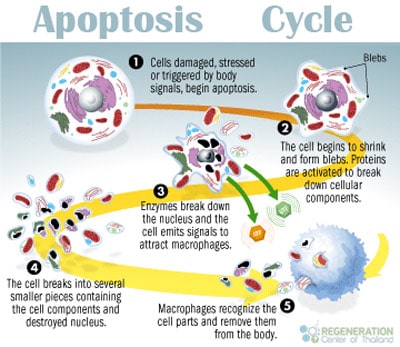
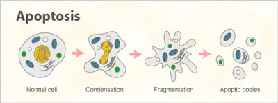
Cells can also be induced/naturally programmed to commit suicide for normal cell turnover and to allow new healthy cells to flourish. The PCD phase starts but the cells shrinking and developing tiny bubble like markers on the surface. After that the DNA & Proteins (chromatin)in the cell nucleus degrades enough to release cytochrome c that breaks down the cells further until the release of UTP and ATP.
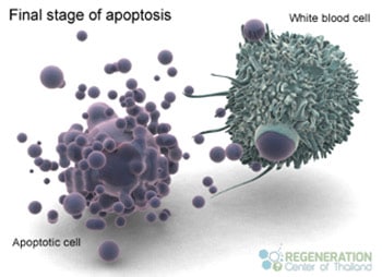
These ATP and UTP nucleotides then bind to the receptors of phagocyte cells such (dendritic cells and macrophages) to help attract themselves to the dying cell. Phospholipid phosphatidylserine then gets exposed to surface creating an “eat me” signal on the phagocytes that devour the remaining cell fragments. These phagocyte cells slowly secrete cytokines IL-10 and TGF-β which inhibits inflammation.
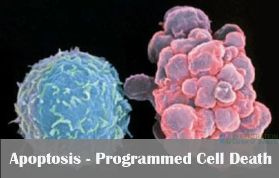
The series of linear events that occur in the suicidal death is so orderly that scientists refer to the process as PCD or programmed cell death. The function of programmed cell death is as intrinsic to our cells the process of mitosis or meiosis. The cycle of a programmed cell death is known as cell apoptosis. (pronounced APE oh TOE sis)
Why do cells commit suicide?
Cells commit suicide for two different reasons.
- Programmed cell death or PCD is needed for natural development. A good example is the formation of our toes and fingers in the fetus that requires nature to use apoptosis to remove some tissue between the fingers and toes to allow for proper function. Another example is in the formation of synapses (signals) between neural cells that require any excess or surplus cells get eliminated through the process of apoptosis.
- PCD is also needed to naturally destroy cells if the body recognizes them as a threat to our system or to maintain the overall integrity of an organism. A good example of Apoptosis is when our cells get infected with a virus. Our body will choose to kill the damaged cells and save the surrounding tissues.
It is believed that defects in our apoptotic process is associated with certain autoimmune conditions such as crohn’s disease, lupus and/or rheumatoid arthritis. Other occurrences of Apoptosis occurs in Cells with DNA or genome damage that may cause disruption in proper embryonic development thus leading the child to birth defects or to stop from becoming cancerous.
Some types of radiation and chemicals that are used in cancer therapies also help to induce apoptosis for some types of cancer cells in the hopes of containment.The survival of our cells requires that it receive constant positive stimulation from surrounding cells. Some well known examples of positive signalling comes from growth factors for neuron cells and Interleukin-2 which is an essential factor needed for mitosis of lymphocytes
On the other side of the spectrum,the receipt of negative signals from cells can also lead to apoptosis of sensory motor neurons. Increasing levels of oxidants inside the cells can cause damage to our DNA.[1] Other negative signals include:
- X-rays
- Ultraviolet lights “UV”
- Chemotherapy drugs
- Rapid Accumulation of proteins
When these negative signals occur, molecules will begin to bind to the cell surface receptors and natural signal the cells to begin the apoptosis cycle.
Examples of cell death activators include:
- Lymphotoxin or TNF-β
- Tumor necrosis factor-alpha or TNF-α
- Fas ligand or FasL
The 3 Primary Mechanisms of Cell Apoptosis
A Cell commits suicide by apoptosis using 3 types of mechanisms:
- Internal signals (mitochondrial pathway)
- External signals (death receptor pathway)
- Apoptosis-Inducing Factor or AIF [2]
Cancer and Apoptosis
Some viruses that are associated with cancer can use biological trickery to prevent the natural cycle of apoptosis in the cells they have invaded. Examples include the human papilloma viruses or HPV that has been known to cause cervical cancer. The Epstein-Barr Virus or EBV is another example of cell trickery and is associated with some types of lymphomas and can also cause mononucleosis.
Apoptosis in our Immune System
The primary function of our immune system us to respond and protect our bodies from foreign invaders through the proliferation of lymphocyte cells (T and/or B cells). After our systems have been protected and the job is done, the cells must be removed to allow the healing process. This removal of cells is done through the process of apoptosis.
Apoptosis Vs Necrosis
The opposite of apoptotic cell death is known as cell necrosis. Necrosis is considered a toxic process in our bodies where the cells become passive victims. Necrosis generally refers to the natural or “un-programmed” degradative processes that occurs after cell death. Many scientists do not like to use the term necrosis to describe cell death.[3]
Oncosis the term that is used to describe the process of events that lead to necrosis. In necrosis, cell swelling and karyolysis occur compared to apoptosis which causes death through pyknosis, karyorrhexis and cell shrinkage. The medical terms “oncotic necrosis” or “oncotic cell death” are sometimes used to describe programmed cell death that is followed up by by cell swelling,[4]
Natures ability to control the life & death of cells has tremendous therapeutic potential in regenerative medicine. Its crucial that we understand when Inappropriate apoptosis occurs as it plays a vital factor in many medical conditions such as neurodegenerative brain diseases such as ALS,Motor Neuron Disease, ischemic heart disease, cancers and many types of autoimmune disorders. The stem cell regeneration center will continue to publish the latest findings, advancements and clinical trials to the benefit all patients across the world.
To learn more about our Apoptosis and how programmed cell death plays a role in medical treatments and therapies please contact us today.
Published Clinical Citations
[1] ^ Ekchariyawat, Peeraya, Arunee Thitithanyanont, Stitaya Sirisinha, and Pongsak Utaisincharoen. 2013. Involvement of GRIM-19 in apoptosis induced in H5N1 virus-infected human macrophages. Innate immunity, no. 6 (March 25). doi:10.1177/1753425913479149. https://www.ncbi.nlm.nih.gov/pubmed/23529854
[2] ^ Kheansaard, Wasinee, Prapaporn Panichob, Suthat Fucharoen, and Dalina I Tanyong. 2011. Cytokine-induced apoptosis of beta-thalassemia/hemoglobin E erythroid progenitor cells via nitric oxide-mediated process in vitro. Acta haematologica, no. 4 (September 21). doi:10.1159/000329903. https://www.ncbi.nlm.nih.gov/pubmed/21934298
>
[3] ^ Kumar, Sunil, Anup Singh Pathania, A K Saxena, R A Vishwakarma, Asif Ali, and Shashi Bhushan. 2013. The anticancer potential of flavonoids isolated from the stem bark of Erythrina suberosa through induction of apoptosis and inhibition of STAT signaling pathway in human leukemia HL-60 cells. Chemico-biological interactions, no. 2 (July 9). doi:10.1016/j.cbi.2013.06.020. https://www.ncbi.nlm.nih.gov/pubmed/23850732
[4] ^ Yorsangsukkamol, Juthaporn, Angkana Chaiprasert, Tanapat Palaga, Therdsak Prammananan, Kiatichai Faksri, Prasit Palittapongarnpim, and Narapon Prayoonwiwat. 2011. Apoptosis, production of MMP9, VEGF, TNF-alpha and intracellular growth of M. tuberculosis for different genotypes and different pks5/1 genes. Asian Pacific journal of allergy and immunology, no. 3. https://www.ncbi.nlm.nih.gov/pubmed/22053594
<